Back
Poster, Podium & Video Sessions
Podium
PD38: Urodynamics/Lower Urinary Tract Dysfunction/Female Pelvic Medicine: Non-neurogenic Voiding Dysfunction II
PD38-10: Upright Open MRI with standing Yoga poses indicates that postural effects on the bladder neck and urethra occur relevant to female urinary incontinence.
Sunday, May 15, 2022
8:30 AM – 8:40 AM
Location: Room 244
Lynn stothers*, Andrew Macnab, vancouver, Canada
- LS
Lynn Stothers, MD
University of California Los Angeles
Podium Presenter(s)
Introduction: The practice of Yoga including physical postures and breath work has been studied in the treatment of urinary incontinence (UI) in women. The open configuration of the magnet in upright MRI systems (MRO) now allows studies in standing subjects.
Objective: to develop an imaging protocol incorporating Yoga poses to evaluate changes in lower urinary tract (LUT) anatomy affecting the bladder neck and urethra that result from alternations in posture.
Methods: Female cases (UI > 1 year, surgically naïve) and controls (No UI or LUTS on ICIQ) completed an MRO imaging protocol using a 0.5 T Paramed scanner. Following an iterative design process, the protocol included imaging sequences for 8 Yoga poses; trikonasana (triangle pose), Utkatasana (chair pose), virabhadrasana 2 (warrior 2 pose), squat (malasana), supine supported bridge and legs up the wall. Each subject acted as their own control using Savasana pose as baseline. Sagittal, coronal, and transverse images were obtained in 2 – 5 minute sequences for each pose (MRO technical protocol: Field of view 30, Interslice space 1 mm, slice thickness 2 mm) with oblique orientation to facilitate urethral 3D reconstruction.
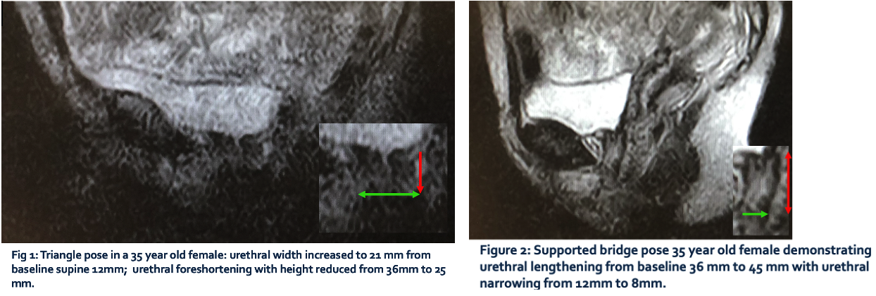
Results: N=12 studies; 3 controls; ages 22-59. Average imaging time required was 2 minutes 42 second per pose. Changes in the urethral length, width and overall structural shape were evident in individuals when comparing supine to upright postures. An upright position with leg abduction demonstrated the highest degree of urethral widening (from baseline 12 mm (+/-3) to 21 mm (+/- 4)) and foreshortening (from baseline 36 mm (+/-3) to 25 mm (+/-2)) (fig 1). Supine bridge position uniquely narrowed the urethra (baseline 12 mm (+/- 3) to 8 mm (+/- 3)), increasing length (baseline 36mm (+/- 3) to 45 mm (+/- 2)) of the urethra compared to standing posture where the bladder neck region widened (fig 2).
Conclusions: Open MRI demonstrates that changes in urethral anatomy occur when upright and supine Yoga postures are compared. Importantly, this protocol proved feasible for subjects to complete, and provided images with sufficient resolution to allow visualization of the postural effects on the bladder neck and urethra. Future studies will enable more detailed anatomic effects of posture, gravity and position on the LUT to be identified, and likely provide insights into postural effects of relevance to female incontinence.
Source of Funding: N/A


.jpg)
.jpg)