Back
Poster, Podium & Video Sessions
Podium
PD39: Benign Prostatic Hyperplasia: Epidemiology, Evaluation & Medical Non-surgical Therapy
PD39-05: Prostate Measurement on Computed Tomography: Variance Decreases as Prostate Volume Increases
Sunday, May 15, 2022
7:40 AM – 7:50 AM
Location: Room 243
Anastasija Useva, Bethany Regan, Michael Iorga*, Alexandr Pinkhasov, Timothy Byler, Scott Wiener, Syracuse, NY
- MI
Podium Presenter(s)
Introduction: Given that yearly prostate growth is minimal in benign prostatic hyperplasia (BPH), there has been increasing reliance on prior imaging to measure prostate volume. Magnetic resonance imaging (MRI) planimetry, transrectal ultrasound (TRUS), and specimen weight after radical prostatectomy correlate highly. However, previous studies report that computed tomography (CT) overestimates volumes of small prostates and underestimates volumes of large prostates. These studies have been limited to prostate sizes less than 60 grams. We evaluated the degree of agreement between CT and MRI in different prostate sizes and evaluated the role of observer bias.
Methods: A retrospective review of 700 men with BPH who had undergone MRI fusion biopsy was performed. Men with available CT scans – with or without contrast – within two years of their MRI were included. Those with indwelling catheters, history of prostate intervention, or extracapsular extension on MRI were excluded. Two medical students, urology residents, and urology attendings repeated three oblate sphere measurements using CT. Our reference was prostate size by MRI planimetry. Bland-Altman analysis was used to determine degree of agreement between CT and MRI. R2 values were used to assess variance of inter-observer measurement based on prostate size and reviewer level of training.
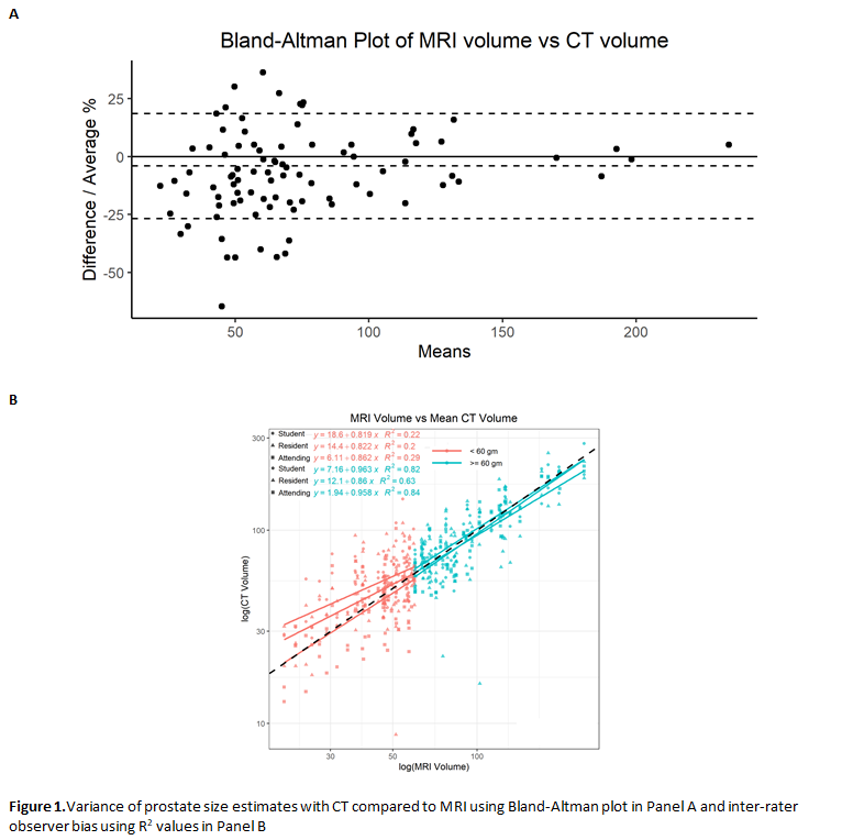
Results: There were 89 men who met inclusion criteria, resulting in 1602 volume measurements. They were grouped as =60gm or <60g, based on the median size of 59.7gm. Mean age was significantly higher in the =60gm than the <60gm group (69.5 vs 65, p=0.004). Race, BMI, and presence of prostate cancer were not significantly different between groups. Figure 1A shows a relative decrease in observer bias between CT and MRI volumes as prostate size increases. Figure 1B shows the degree of inter-rater variance between CT and MRI volumes in the two size groups. R2 values for students, residents, and attendings were 0.22, 0.2, and 0.29 respectively for small glands and 0.82, 0.63, and 0.84 respectively for large glands.
Conclusions: Compared to MRI planimetry, CT accurately assesses prostate size for glands =60gm and overestimates prostate size for glands <60gm. This may be attributable to inadvertent inclusion of peri-prostatic connective tissues which represent a larger fraction of total gland volume.
Source of Funding: Endourological Society


.jpg)
.jpg)