Back
Poster, Podium & Video Sessions
Video
V03: Urolithasis/Endourology
V03-06: Ectopic Pyelolithotomy via a Combined Robotic and Endoscopic Approach
Friday, May 13, 2022
1:50 PM – 2:00 PM
Location: Video Abstracts Theater
Andrew Higgins*, Alex Nourian, Joshua Cohn, Justin Friedlander, Philadelphia, PA

Andrew Higgins, MD
Einstein Medical Center, Philadelphia
Video Presenter(s)
Introduction: Ectopic kidneys are rare congenital anomalies and may be susceptible to higher rates of nephrolithiasis, reflux, and ureteropelvic junction obstruction due to their different orientation. Treatment of nephrolithiasis in patients with pelvic kidneys remains challenging due to the complex anatomy and surrounding structures. We describe the combined robotic and endoscopic treatment of a patient with an ectopic left kidney with a large kidney stone burden.
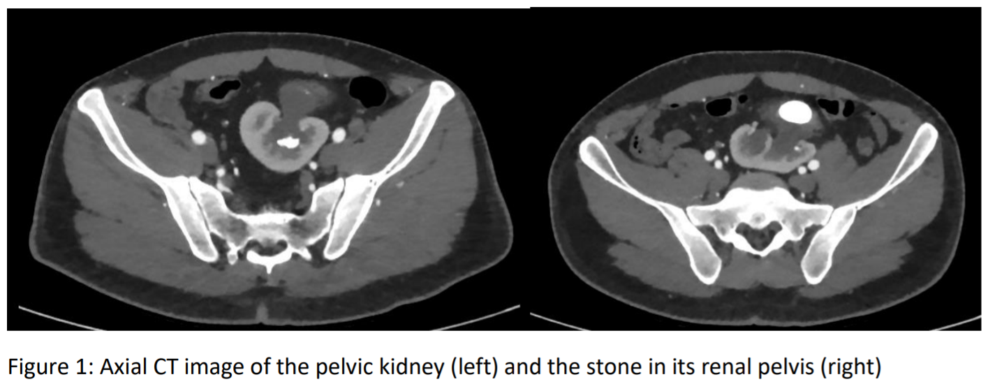
Methods: A 35-year-old male presented with abdominal pain was found to have a pelvic kidney with a 3.1 cm staghorn calculus with mild hydronephrosis, as well as additional lower pole renal stones measuring up to 1.3 cm (Figure 1). Preoperative computed tomography angiography demonstrated that the ectopic kidney was anterior to the bifurcation of the iliac vessels with overlying bowel, thus making a percutaneous approach challenging; however, the anterior orientation of the renal pelvis appeared amenable to combined robotic-assisted laparoscopic and endoscopic pyelolithotomy.
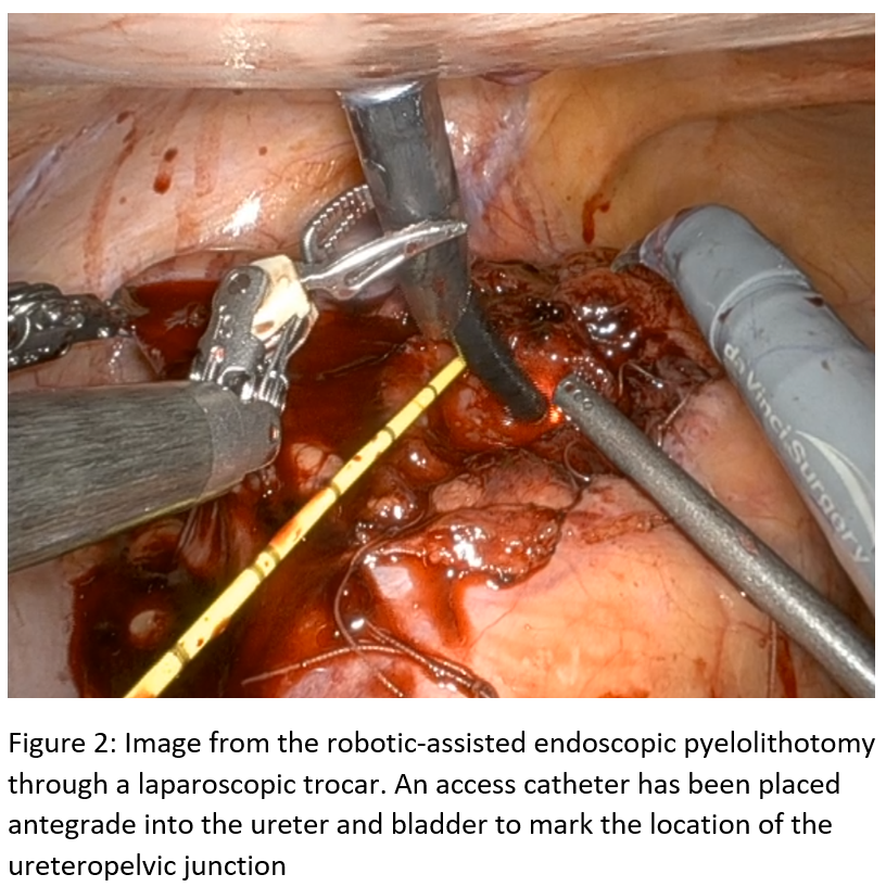
Results: The kidney was accessed via a traditional pelvic surgery robotic port set up. The renal pelvis was sharply incised to deliver the staghorn calculus in one specimen. A flexible cystoscope was introduced via a laparoscopic trocar and guided into the incised renal pelvis and into the lower pole to remove remaining stones via basket extraction and aspiration via the laparoscopic suction-irrigator. A ureteral stent was advanced through the pyelotomy and into the bladder via a guidewire before watertight closure of the renal pelvis and reapproximation of the peri-renal fat and peritoneum. The patient was discharged on postoperative day one following removal of his foley catheter and Jackson-Pratt drain. He underwent an uneventful stent removal in the office 6 weeks postoperatively.
Conclusions: Combined robotic-assisted and endoscopic pyelolithotomy can be a safe and effective approach for pelvic kidney stone cases that may not be amenable to a single treatment modality.
Source of Funding: none


.jpg)
.jpg)