Resident Department of Periodontology, Tokyo Dental College
Aim: In our previous study, the use of FGF-2 with DBBM was similarly effective as FGF-2 alone in the healing of experimental periodontal defects in rats. It was suggested that the combined use of FGF-2 and DBBM enhances healing via promotion of cell proliferation, angiogenesis, and osteogenic differentiation. Combined use of signaling molecules and bone substitutes is clinically recommended for the treatment of poorly-contained defects. The purpose of this study is to investigate the effects of the combined use of FGF-2 and DBBM on the periodontal healing of poorly-contained defects in rats. Materials and
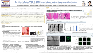
Methods: Poorly-contained (2-wall) periodontal defects were created mesially to the maxillary first molars in Wistar rats. Defects were filled with FGF-2, DBBM, FGF-2+DBBM, or left unfilled. Histological examinations and microcomputed tomography were used to evaluate healing at 4 weeks postoperatively. In vitro, the positive VEGF expression of rat bone marrow mesenchymal stem cells (BMSCs) after 72 h of seeding on DBBM with/without FGF-2 was assessed by confocal laser scanning microscopy (CLSM).
Results: Histologically, greater bone formation was observed in FGF-2 and FGF-2+DBBM groups than in Unfilled or DBBM groups. Bone volume fraction was significantly greater in FGF-2+DBBM groups than that in Unfilled group. In vitro, CLSM images revealed that the positive staining for VEGF in BMSCs was more frequently observed in FGF-2+DBBM group compared with that in DBBM group.
Conclusions: It was suggested that DBBM acted as a scaffold, spacemaker, and carrier of FGF-2, and thereby accelerates healing of poorly-contained defects in FGF-2+DBBM group.