Back
abs: Novel 3D Tumor Model for Surgical Education
Small Animal
abs: Novel 3D Tumor Model for Surgical Education
Innovations in Surgical Oncology
Saturday, October 15, 2022
3:30pm – 3:45pm
Location: Oregon Ballroom 202

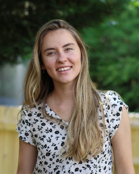
Abby Cox Laws
NC State College of Veterinary Medicine
Cary, North Carolina
Abstract Presenter(s)
Alternative laboratory teaching methods are becoming increasingly desirable and effective in medical education environments. While there are ethical concerns around using cadaveric models, the COVID-19 pandemic created an emergent need for teaching tools that can be used off-site, and adequately convey surgical principles and replicate conventional on-site practical training. We have developed a 3-dimensional custom-made silicone soft tissue tumor model using 3D-printed molds derived from canine soft tissue sarcoma computed tomography images. We hypothesized that this model would be representative of the pathologic environment and different tissue layers encountered during tumor excision, producible in high numbers, and effective in teaching surgical oncology principles. A large cohort of students in addition to a small number of professional veterinarians at different levels of specialty training followed the laboratory guidelines and evaluated the simulated tumor model based on a survey. All participants were able to successfully complete the practical training. The model also allowed the students to identify and correct technical errors associated with biopsy sampling and margin dissection, and to understand the clinical impacts related to those errors. Face and content validity of the model were assessed using Likert-style questionnaires with expert scores of 3.8/5 and 4.3/5, respectively. Although limited by the difficulty of replicating the physical properties of biological tissue, this model development emphasizes the efficacy of alternative non-cadaveric laboratory teaching tools and could become a valuable aid in the veterinary curricula.
