Back
Abstract Session
Genetics, genomics and proteomics
Session: Abstracts: Genetics, Genomics and Proteomics (0562–0565)
0565: Deciphering Complement System-dependent Cellular Pathways in Human Rheumatoid Arthritis Synovial Tissues Using Single-cell Computational Omics
Sunday, November 13, 2022
8:45 AM – 8:55 AM Eastern Time
Location: Room 120

Fan Zhang, PhD
The University of Colorado
Aurora, CO, United States
Presenting Author(s)
Juan Vargas1, Nirmal Banda2, Ian Mantel3, Anna Jonsson4, Kevin Wei5, Deepak Rao4, Susan Goodman6, Kevin D Deane7, Jennifer Seifert8, Jennifer Anolik9, Michael Brenner10, Soumya Raychaudhuri4, Accelerating Medicines Partnership RA/SLE4, Michael Holers2, Laura Donlin6 and Fan Zhang8, 1School Public Health Biostatistics Department, Aurora, CO, 2Department of Medicine Division of Rheumatology, Aurora, CO, 3Weill Cornell Medicine, New York, NY, 4Brigham and Women's Hospital, Boston, MA, 5Brigham and Women's Hospital and Harvard Medical School, Boston, MA, 6Hospital for Special Surgery, New York, NY, 7University of Colorado Denver Anschutz Medical Campus, Denver, CO, 8University of Colorado, Aurora, CO, 9University of Rochester Medical Center, Rochester, NY, 10Brigham and Women's Hospital, Harvard Medical School, Boston, MA
Background/Purpose: The complement system is a major component of innate immunity and plays a vital role in experimental models of autoimmune arthritis pathogenesis. In patients with RA, local activations of the complement pathways in the synovium will interact with macrophages as well as other synovial mesenchymal cells that cause tissue inflammation. However, it remains to be determined how specific complement components in the RA synovium modulate cell type functions to both enhance injury but also strengthen phagocytosis and inflammation resolution.
Methods: AMP RA/SLE has generated a comprehensive synovial single-cell multimodal cell atlas ( >314,000 cells, 70 RA patients) and classified the spectrum of RA biopsies into six groups named CTAPs (Cell Type Abundance Phenotypes)1. In this study, we link complement activation pathways with cell-type heterogeneity, and test associations of complement components across different CTAPs using Generalized Linear Mixed Models. Furthermore, we characterize myeloid cell differentiation using single-cell trajectory analysis and align specific complement pathways to myeloid functional polarization states. Finally, we performed cell type interactome analysis to reveal complement-dependent receptor-ligand pairs among multiple cell types that likely contribute to tissue inflammation.
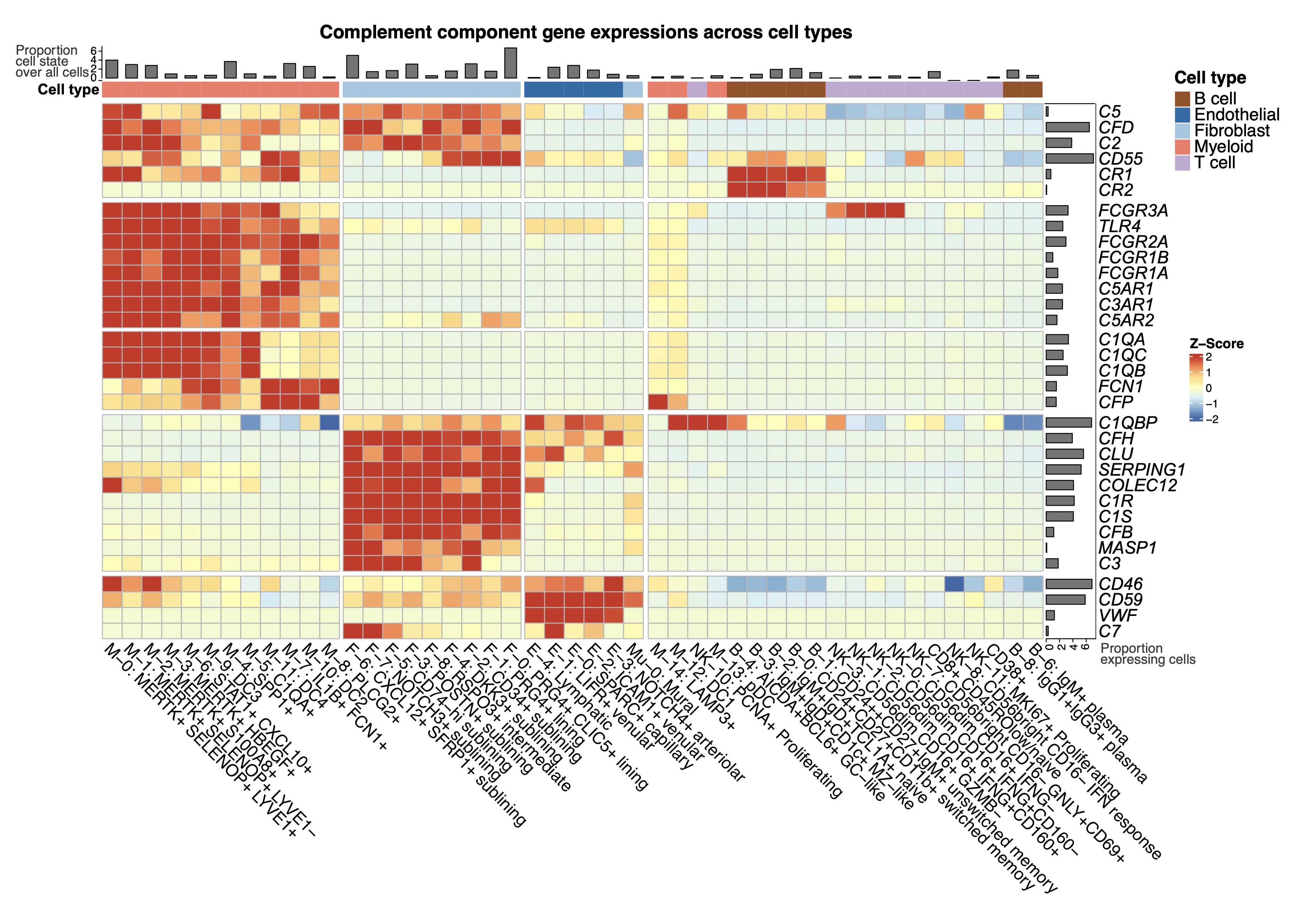
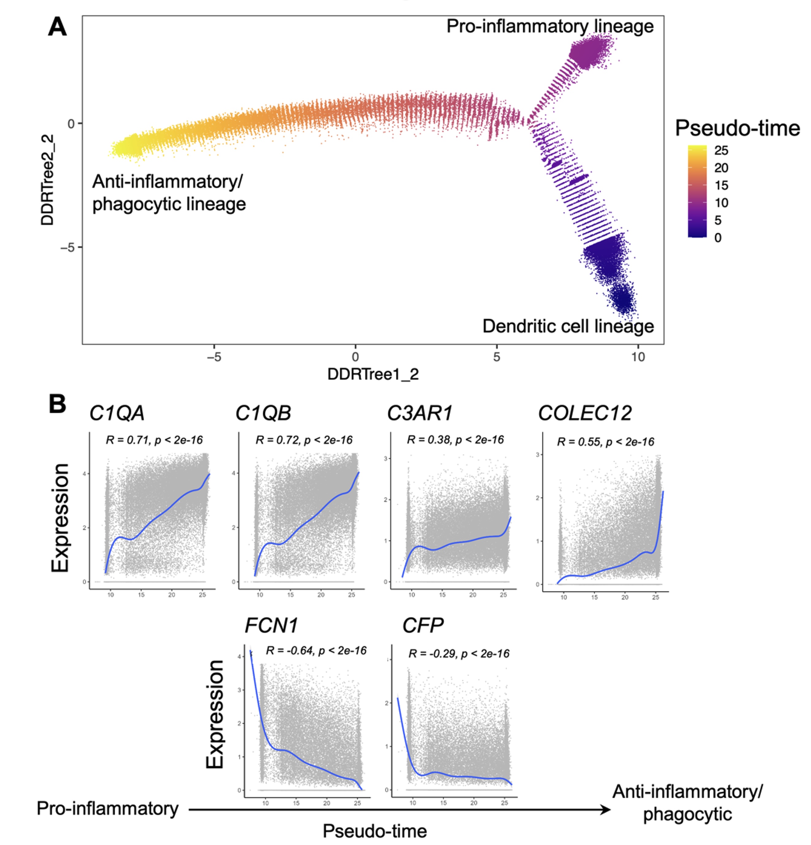
Results: We generated an Autoimmune Complement Cellular Graph (ACCG) comprehensively characterizing interactions between complement components and cell-type co-varying patterns in RA synovium. We found that complement components present distinct transcriptomic patterns across cell types in unexpected patterns (Fig. 1). Furthermore, each synovial CTAP contains a distinct selection of myeloid cell states. Specifically, expressions of complement C1QA-C, C3AR1, and COLEC12 are correlated with myeloid phagocytic trajectory, while complement FCN1 and CFP are correlated with myeloid pro-inflammatory trajectory, which suggests different complement pathways interact with myeloid states in RA synovium (Fig. 2). Intriguingly, the phagocytic MERTK+ tissue macrophages in CTAP-EFM (dominated by myeloid, fibroblast, and endothelial cells) display two complement pathways, namely the phagocytic C1QA-C factors and the inflammatory receptor C3AR1, while sublining fibroblasts highly expressed complement C3, which suggests the potential interactions between MERTK+ macrophages and specific fibroblasts that activate complement pathways in the synovium.
Conclusion: Through systematically aligning complement pathways with synovial cellular heterogeneity, we generated a map describing how specific complement components can modulate functional myeloid states to strengthen resolution in RA patients through phagocytic functions. This approach of analyzing high-resolution single-cell multimodal data in human tissues with a focus on the complement system brings insights into new complement pathway-based cellular targets for patients with RA, and provides a roadmap for similar studies of other complex pathways in tissues.
 Fig. 1. Single-cell expression distributions of complement components across cell types in rheumatoid arthritis synovial tissues. Different myeloid, fibroblast and endothelial cell states express different complement components.
 Fig. 2. Myeloid-specific single-cell trajectory analysis. A. Identification of trajectories for myeloid cell differentiation: pro-inflammatory lineage, anti-inflammatory lineage, and dendritic cell lineage, B. Complement components C1QA-B, C3AR1, and COLEC12 expressions correlated with anti-inflammatory/phagocytic function, and complement components FCN1 and CFP expressions correlated with pro-inflammatory function. Spearman correlation R and p-value are shown.
Disclosures: J. Vargas, None; N. Banda, None; I. Mantel, None; A. Jonsson, Amgen; K. Wei, Gilead sciences, Mestag, nanoString, 10X Genomics; D. Rao, Janssen, Merck, Bristol-Myers Squibb, Scipher Medicine, HiFiBio, Inc., AstraZeneca, Pfizer; S. Goodman, Novartis, UCB; K. Deane, Werfen; J. Seifert, None; J. Anolik, None; M. Brenner, GSK, 4FO Ventures, Mestag Therapeutics; S. Raychaudhuri, Mestag, Inc, Rheos Medicines, Janssen, Pfizer, Biogen; A. RA/SLE, None; M. Holers, Celgene, Bristol-Myers Squibb(BMS), Janssen; L. Donlin, None; F. Zhang, None.
Background/Purpose: The complement system is a major component of innate immunity and plays a vital role in experimental models of autoimmune arthritis pathogenesis. In patients with RA, local activations of the complement pathways in the synovium will interact with macrophages as well as other synovial mesenchymal cells that cause tissue inflammation. However, it remains to be determined how specific complement components in the RA synovium modulate cell type functions to both enhance injury but also strengthen phagocytosis and inflammation resolution.
Methods: AMP RA/SLE has generated a comprehensive synovial single-cell multimodal cell atlas ( >314,000 cells, 70 RA patients) and classified the spectrum of RA biopsies into six groups named CTAPs (Cell Type Abundance Phenotypes)1. In this study, we link complement activation pathways with cell-type heterogeneity, and test associations of complement components across different CTAPs using Generalized Linear Mixed Models. Furthermore, we characterize myeloid cell differentiation using single-cell trajectory analysis and align specific complement pathways to myeloid functional polarization states. Finally, we performed cell type interactome analysis to reveal complement-dependent receptor-ligand pairs among multiple cell types that likely contribute to tissue inflammation.
Results: We generated an Autoimmune Complement Cellular Graph (ACCG) comprehensively characterizing interactions between complement components and cell-type co-varying patterns in RA synovium. We found that complement components present distinct transcriptomic patterns across cell types in unexpected patterns (Fig. 1). Furthermore, each synovial CTAP contains a distinct selection of myeloid cell states. Specifically, expressions of complement C1QA-C, C3AR1, and COLEC12 are correlated with myeloid phagocytic trajectory, while complement FCN1 and CFP are correlated with myeloid pro-inflammatory trajectory, which suggests different complement pathways interact with myeloid states in RA synovium (Fig. 2). Intriguingly, the phagocytic MERTK+ tissue macrophages in CTAP-EFM (dominated by myeloid, fibroblast, and endothelial cells) display two complement pathways, namely the phagocytic C1QA-C factors and the inflammatory receptor C3AR1, while sublining fibroblasts highly expressed complement C3, which suggests the potential interactions between MERTK+ macrophages and specific fibroblasts that activate complement pathways in the synovium.
Conclusion: Through systematically aligning complement pathways with synovial cellular heterogeneity, we generated a map describing how specific complement components can modulate functional myeloid states to strengthen resolution in RA patients through phagocytic functions. This approach of analyzing high-resolution single-cell multimodal data in human tissues with a focus on the complement system brings insights into new complement pathway-based cellular targets for patients with RA, and provides a roadmap for similar studies of other complex pathways in tissues.
 Fig. 1. Single-cell expression distributions of complement components across cell types in rheumatoid arthritis synovial tissues. Different myeloid, fibroblast and endothelial cell states express different complement components.
 Fig. 2. Myeloid-specific single-cell trajectory analysis. A. Identification of trajectories for myeloid cell differentiation: pro-inflammatory lineage, anti-inflammatory lineage, and dendritic cell lineage, B. Complement components C1QA-B, C3AR1, and COLEC12 expressions correlated with anti-inflammatory/phagocytic function, and complement components FCN1 and CFP expressions correlated with pro-inflammatory function. Spearman correlation R and p-value are shown.
Disclosures: J. Vargas, None; N. Banda, None; I. Mantel, None; A. Jonsson, Amgen; K. Wei, Gilead sciences, Mestag, nanoString, 10X Genomics; D. Rao, Janssen, Merck, Bristol-Myers Squibb, Scipher Medicine, HiFiBio, Inc., AstraZeneca, Pfizer; S. Goodman, Novartis, UCB; K. Deane, Werfen; J. Seifert, None; J. Anolik, None; M. Brenner, GSK, 4FO Ventures, Mestag Therapeutics; S. Raychaudhuri, Mestag, Inc, Rheos Medicines, Janssen, Pfizer, Biogen; A. RA/SLE, None; M. Holers, Celgene, Bristol-Myers Squibb(BMS), Janssen; L. Donlin, None; F. Zhang, None.

