Back
Poster Session D
Crystal arthropathies
Session: (1787–1829) Metabolic and Crystal Arthropathies – Basic and Clinical Science Poster
1800: Successful Establishment of Chronic Gouty Arthritis Model in C57BL/6 Mice
Monday, November 14, 2022
1:00 PM – 3:00 PM Eastern Time
Location: Virtual Poster Hall
- YY
Yue Yin, MD, PhD
Peking Union Medical College Hospital
Beijing, Beijing, China
Abstract Poster Presenter(s)
Yue Yin1, Yun Zhang1, Hong Di1, Xinxin Han1 and Xuejun Zeng2, 1Peking Union Medical College Hospital, Beijing, China, 2Department of General Internal Medicine, Peking Union Medical College Hospital, Chinese Academy of Medical Science & Peking Union Medical College, Beijing, China
Background/Purpose: Gout is an inflammatory disease caused by the deposition of MSU crystals in joints and other parts. At present, little progress in the research of chronic gouty arthritis (CGA) has been reported, which is featured by recurrent arthritis and formation of tophus. In order to further explore the mechanisms of gout, the animal models of gouty arthritis are needed. By far, there have been attempts to establish acute gouty arthritis (AGA) model by direct injection of MSU crystal. However, there are still difficulties in the establishment of CGA model in mice. Therefore, we further explored the method of establishing CGA mice model on the basis of the existing AGA mice model.
Methods: According to the previous literature reports, 8-week-old C57BL / 6J mice were injected with MSU crystal suspension (model group) / PBS (control group) into their hind paws to establish a mouse model of AGA. Joint swelling was expressed as the ratio of the footpad thickness of the inflamed over the normal control joint. Based on the AGA model, the repeated acute inflammation and remission process of chronic gouty arthritis was established by repeatedly injecting MSU crystal into the footpads for up to 4 months. The tophi in joints were observed, imaging and pathological examination were conducted. The mice in CGA group were sacrificed seven days after injection of MSU crystal for the last time. Then the bone destruction in pathology and the expression of MMP3 and RANKL were evaluated. The uric acid level in AGA and CGA model was also detected.
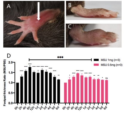
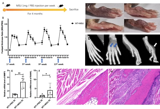
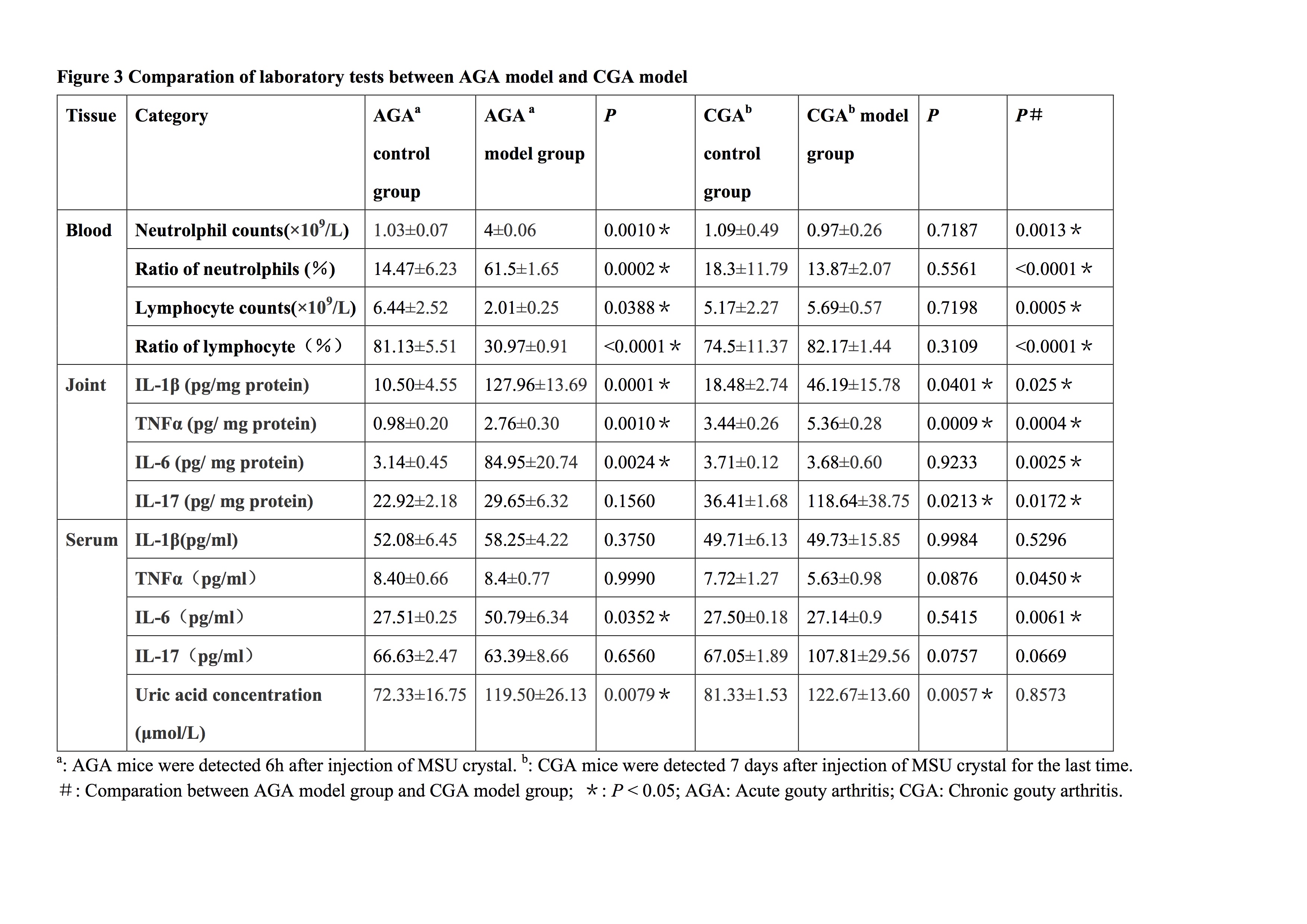
Results: In AGA model, there was significant swelling of joints (Figure 1A-C), which was time and concentration dependent. (Figure 1D). Based on the results of AGA model, the CGA model was successfully established (Figure 2A). The repeated acute inflammation and remission process were seen in joints of CGA model (Figure 2B), and tophus was observed on the surface of the footpad (Figure 2C). Imaging showed bone deformity and abnormal material deposition around the bone (Figure 2D). In CGA model, the mRNA expression levels of MMP3 and RANKL were higher than those in control group (P=0.0152 and 0.0043, respectively) (Figure 2E-F). Pathology showed significant fibrous tissue formation with lymphocytes infiltration, which invaded the bone and led to bone destruction (Figure 2G-H). In the mouse CGA model, IL-1β, TNFα, IL-17 were significantly increased in local joint tissues (P =0.0401, 0.0009, 0.0213, respectively), indicating consistent inflammation in joints of CGA model. In AGA model, the blood uric acid increased rapidly after local injection of MSU crystal, but returned to normal after 12 hours. After repeated injection and formation of tophus in CGA model, the serum uric acid was still in a high level after injection of MSU seven days ago (Figure 3).
Conclusion: We successfully established the mouse experimental model of CGA, and observed the manifestations of recurrent arthritis, formation of tophus, bone destruction, and increase in serum uric acid level. The establishment and application of CGA mice model may be a great help for the study of chronic gout in the future.
 Figure 1. (A-C) The establishment of acute gouty arthritis (AGA) model. (B) AGA control group (injection of PBS to joints). (C) AGA model group (injection of MSU to joints). (D) Changes in footpad thickness ratio in different time and concentration of MSU crystal suspension.
 Figure 2. The establishment of chronic gouty arthritis (CGA) model. (A) Timeline of establishment of CGA models. (B) Footpad thickness ratio in different time of CGA model establishment. (C) Tophi of joints. (D) CT reconstruction of footpads. (E-F) Expression of MMP3 (E) and RANKL (F) in joint of control group (injection of PBS to joints) and AGA model group (injection of MSU to joints). (G-H) Pathology of joint in CGA models. (G) Control group; (H) CGA group.

Disclosures: Y. Yin, None; Y. Zhang, None; H. Di, None; X. Han, None; X. Zeng, None.
Background/Purpose: Gout is an inflammatory disease caused by the deposition of MSU crystals in joints and other parts. At present, little progress in the research of chronic gouty arthritis (CGA) has been reported, which is featured by recurrent arthritis and formation of tophus. In order to further explore the mechanisms of gout, the animal models of gouty arthritis are needed. By far, there have been attempts to establish acute gouty arthritis (AGA) model by direct injection of MSU crystal. However, there are still difficulties in the establishment of CGA model in mice. Therefore, we further explored the method of establishing CGA mice model on the basis of the existing AGA mice model.
Methods: According to the previous literature reports, 8-week-old C57BL / 6J mice were injected with MSU crystal suspension (model group) / PBS (control group) into their hind paws to establish a mouse model of AGA. Joint swelling was expressed as the ratio of the footpad thickness of the inflamed over the normal control joint. Based on the AGA model, the repeated acute inflammation and remission process of chronic gouty arthritis was established by repeatedly injecting MSU crystal into the footpads for up to 4 months. The tophi in joints were observed, imaging and pathological examination were conducted. The mice in CGA group were sacrificed seven days after injection of MSU crystal for the last time. Then the bone destruction in pathology and the expression of MMP3 and RANKL were evaluated. The uric acid level in AGA and CGA model was also detected.
Results: In AGA model, there was significant swelling of joints (Figure 1A-C), which was time and concentration dependent. (Figure 1D). Based on the results of AGA model, the CGA model was successfully established (Figure 2A). The repeated acute inflammation and remission process were seen in joints of CGA model (Figure 2B), and tophus was observed on the surface of the footpad (Figure 2C). Imaging showed bone deformity and abnormal material deposition around the bone (Figure 2D). In CGA model, the mRNA expression levels of MMP3 and RANKL were higher than those in control group (P=0.0152 and 0.0043, respectively) (Figure 2E-F). Pathology showed significant fibrous tissue formation with lymphocytes infiltration, which invaded the bone and led to bone destruction (Figure 2G-H). In the mouse CGA model, IL-1β, TNFα, IL-17 were significantly increased in local joint tissues (P =0.0401, 0.0009, 0.0213, respectively), indicating consistent inflammation in joints of CGA model. In AGA model, the blood uric acid increased rapidly after local injection of MSU crystal, but returned to normal after 12 hours. After repeated injection and formation of tophus in CGA model, the serum uric acid was still in a high level after injection of MSU seven days ago (Figure 3).
Conclusion: We successfully established the mouse experimental model of CGA, and observed the manifestations of recurrent arthritis, formation of tophus, bone destruction, and increase in serum uric acid level. The establishment and application of CGA mice model may be a great help for the study of chronic gout in the future.
 Figure 1. (A-C) The establishment of acute gouty arthritis (AGA) model. (B) AGA control group (injection of PBS to joints). (C) AGA model group (injection of MSU to joints). (D) Changes in footpad thickness ratio in different time and concentration of MSU crystal suspension.
 Figure 2. The establishment of chronic gouty arthritis (CGA) model. (A) Timeline of establishment of CGA models. (B) Footpad thickness ratio in different time of CGA model establishment. (C) Tophi of joints. (D) CT reconstruction of footpads. (E-F) Expression of MMP3 (E) and RANKL (F) in joint of control group (injection of PBS to joints) and AGA model group (injection of MSU to joints). (G-H) Pathology of joint in CGA models. (G) Control group; (H) CGA group.
Disclosures: Y. Yin, None; Y. Zhang, None; H. Di, None; X. Han, None; X. Zeng, None.

