Back
Poster Session B
Antiphospholipid Syndrome
Session: (0671–0694) Antiphospholipid Syndrome Poster
0693: Low Neutrophil Ectonucleotidase Activity Drives Neutrophil-Platelet Aggregation and Platelet Activation in Antiphospholipid Syndrome (APS)
Sunday, November 13, 2022
9:00 AM – 10:30 AM Eastern Time
Location: Virtual Poster Hall
- NS
NaveenKumar Somanathapura, PhD
University of Mysore, Karnataka, India
Ann arbor, MI, United States
Abstract Poster Presenter(s)
Naveen Kumar S K1, Claire Hoy1, Srilakshmi Yalavarthi1, Cyrus Sarosh2, Kaitlyn Sabb1, Christine Rysenga1, Jacqueline Madison1, Yu Zuo1 and Jason S Knight3, 1University of Michigan, Ann Arbor, MI, 2University of Michigan, Temperance, MI, 3University of Michigan, Division of Rheumatology, Ann Arbor, MI
Background/Purpose: By converting extracellular ATP into homeostatic adenosine, the ectonucleotidases CD39 (ATP to AMP) and CD73 (AMP to adenosine) dictate extracellular purinergic gradients and as such are critical players in the regulation of leukocyte and platelet functions. Insufficient activity of these homeostatic ectoenzymes has been associated with neutrophil-driven inflammation. Crosstalk between neutrophils and platelets links thrombotic and inflammatory processes, and elevated levels of neutrophil-platelet aggregates (NPAs) have been reported in various disease states. Understudied are (i) the role that ectonucleotidases play in restraining NPA formation, as well as (ii) the role that NPAs play in the prototypical thromboinflammatory disease, antiphospholipid syndrome (APS). Here, we aimed to study the relationship between the CD39/CD73 axis, neutrophils, and platelets in APS pathophysiology.
Methods: NPAs (CD16+CD15+CD41+) and platelet P-selectin (CD62P) levels were assessed in the blood of patients with durably positive antiphospholipid antibodies (aPL) and features of APS (n=36) by flow cytometry; no subjects had lupus. These 36 subjects were compared to 20 healthy controls. In parallel, neutrophil CD39 and CD73 activities were measured by a standard malachite green assay. Healthy donor neutrophils and platelets (ratio 1:20) were also studied in vitro in the presence or absence of a specific CD39 activity inhibitor (ARL 67156). Potential blockers of NPA formation were also tested including anti-CD62P and anti-CD162 (anti-PSGL-1).
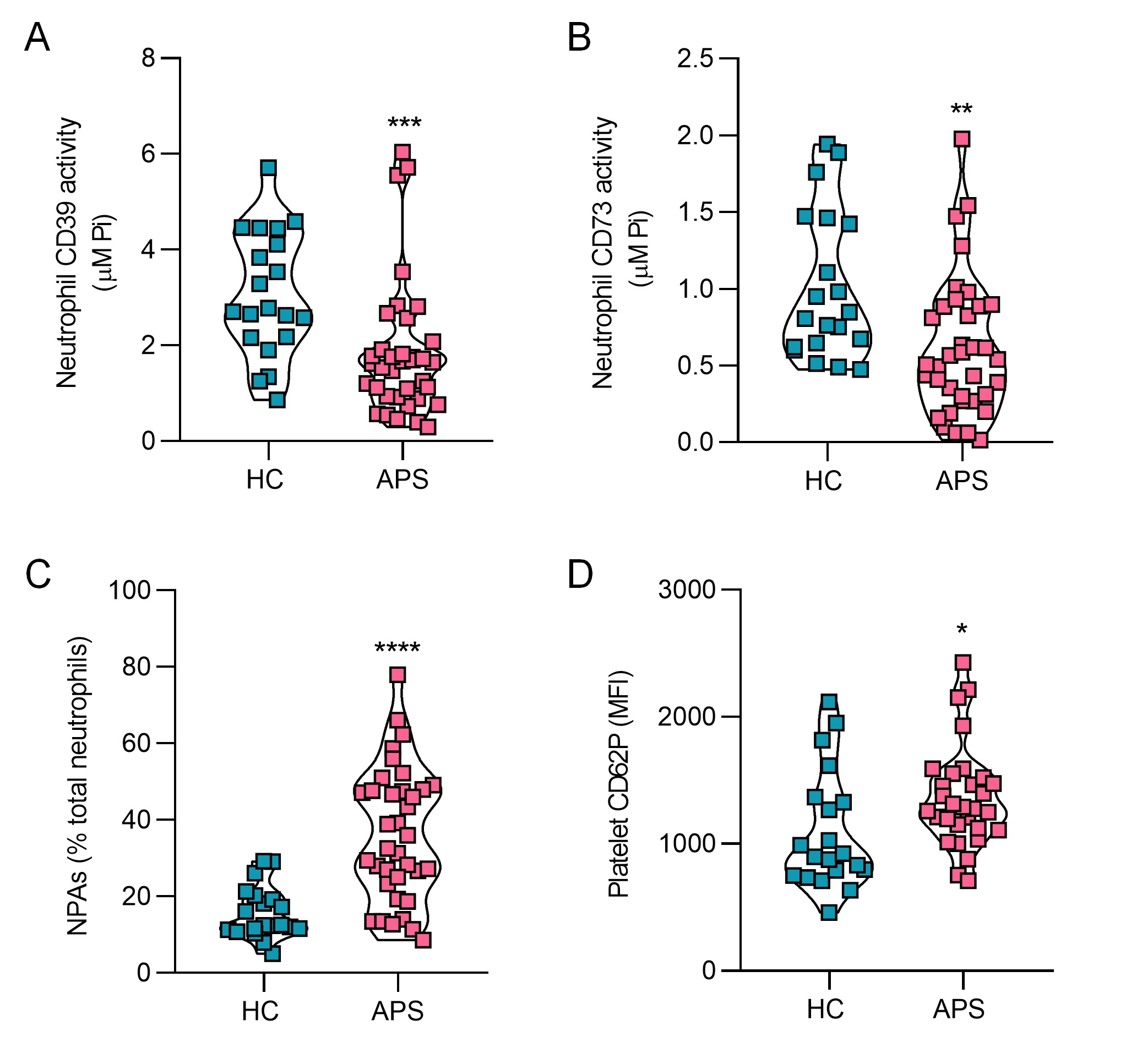
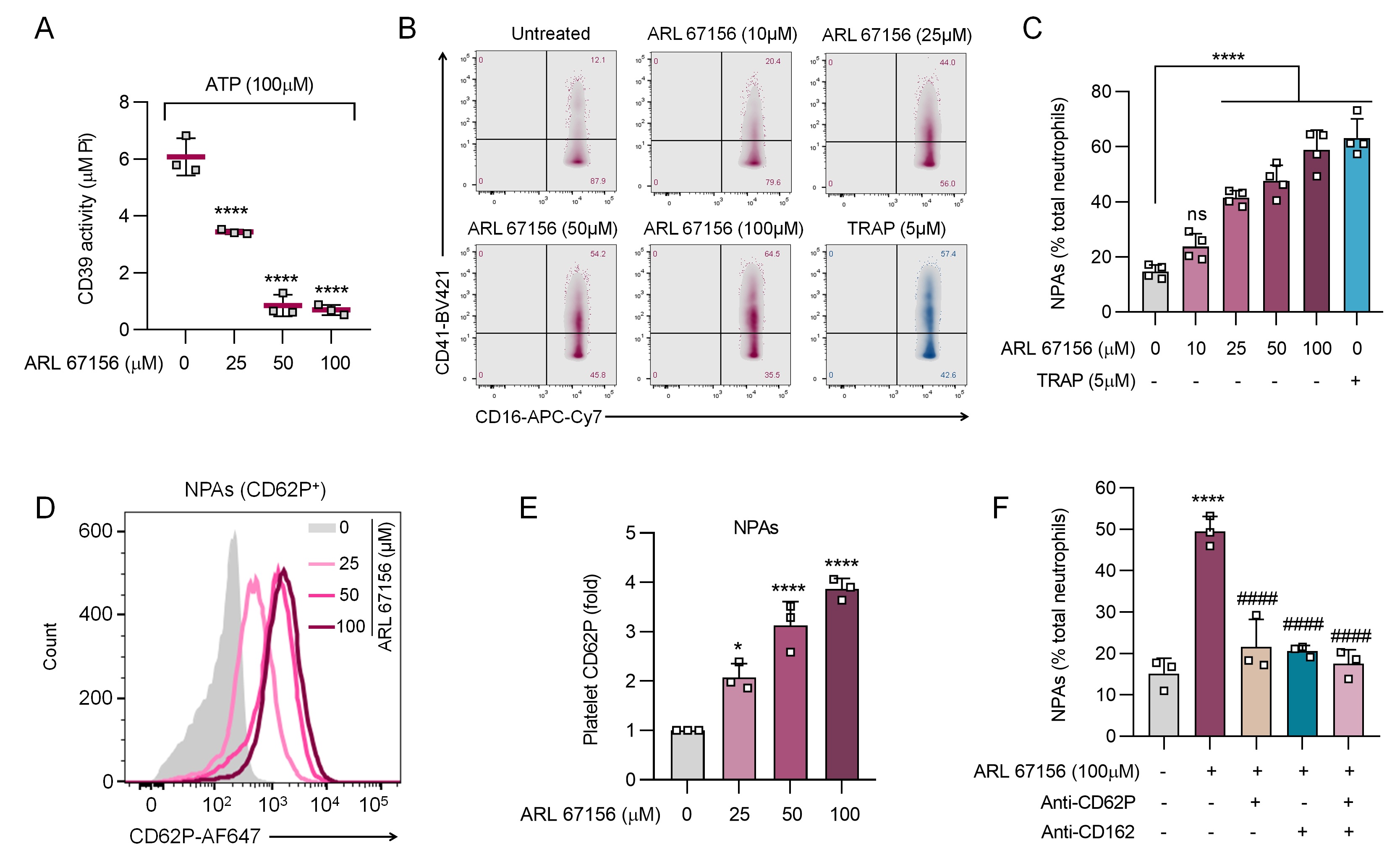
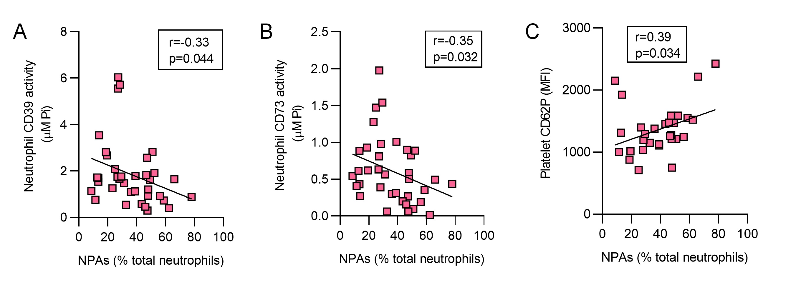
Results: In 36 aPL-positive patients, activities of both neutrophil CD39 (p< 0.001) and neutrophil CD73 (p< 0.001) were markedly decreased as compared with healthy controls (Fig 1A-B). This was paralleled by significant elevations of NPAs (up to 80% of total neutrophils, p< 0.0001) and platelet activation as defined by CD62P expression (p< 0.05, Fig 1C-D). When healthy neutrophils and platelets were cultured together, inhibition of CD39 activity significantly enhanced NPA formation (from baseline 20% to 70%) in dose-dependent fashion (Fig 2A-C). In the same system, increased platelet activation was also observed (2- to 4-fold), as was inhibition of NPA formation by either anti-CD62P or anti-CD162 (both p< 0.0001, Fig 2D-F). In a correlation analysis focused on the aPL-positive patient samples (n=36), circulating NPAs were inversely correlated with neutrophil CD39 activity (r=-0.33, p=0.044) and CD73 activity (r=-0.35, p=0.032, Fig 3A-B). Levels of NPAs also tracked with platelet activation (r=0.38, p=0.034, Fig 3C).
Conclusion: We provide evidence that diminished CD39 and CD73 activity (as appears to be present in many patients with APS) potently potentiate the formation of NPAs. In this context, blocking either CD62P or CD162 significantly tempers NPA formation. Taken together, these findings have potential to identify new approaches to therapy and sub-phenotyping for patients on the APS disease spectrum. Experiments are now underway to determine the upstream regulators of ectonucleotidase activity in APS as well as the downstream pathogenic consequences of excessive NPAs formation.
 Figure 1: Decreased ectonucleotidase activity of APS neutrophils. A-B, CD39 (A) and CD73 (B) activity were measured in neutrophils of 36 patients with positive antiphospholipid antibodies and features of APS and 20 healthy controls (HC). Quantification was by malachite green assay, which measured released inorganic phosphate (Pi) due to ATP (CD39 activity) and AMP (CD73 activity) hydrolysis. C, Assessment of neutrophil-platelet aggregates (NPAs) (CD41+ events within the CD15+CD16+ population) in whole blood. D, Platelet activation as measured by P-selectin (CD62P) expression in whole blood. Groups were compared by Mann-Whitney test; *p < 0.05, **p < 0.01, ***p < 0.001, and ****p < 0.0001.
 Figure 2: CD39 inhibition leads to NPA formation via P-selectin and PSGL-1. A, Determination of CD39 activity in neutrophil-platelet cultures (1:20) in the presence of various concentration of ARL 67156 (CD39 inhibitor). Released inorganic phosphate (Pi) due to ATP hydrolysis was quantified by malachite green assay. B-C, Representative flow cytometric analysis of neutrophil-platelet aggregates (NPAs) showing neutrophils (CD16+CD41-) and NPAs (CD16+CD41+) upon stepwise CD39 inhibition (n=4/group). D-E, Platelet CD62P expression within the NPAs population was assessed using flow cytometry (n=3/group). F, NPA formation in the presence of anti-CD62P and anti-CD162 (anti-PSGL-1) upon CD39 inhibition (n=3/group). Data are presented as mean ± SD; *p < 0.05, **p < 0.01, ***p < 0.001, ****p < 0.0001, and ns=not significant compared with untreated; ####p < 0.0001 compared with the ARL 67156-alone condition. Analysis was by one-way ANOVA followed by Tukey's multiple-comparison test.
 Figure 3: NPAs correlate with low neutrophil CD39 expression, low neutrophil CD73 expression, and platelet activation. A-C, Neutrophil-platelet aggregates (NPAs) correlated with neutrophil CD39 (A) and CD73 (B) activity, as well as platelet CD62P expression (C). Spearman’s correlation coefficients are presented for n=36 patients with positive antiphospholipid antibodies and features of APS.
Disclosures: N. S K, None; C. Hoy, None; S. Yalavarthi, None; C. Sarosh, None; K. Sabb, None; C. Rysenga, None; J. Madison, None; Y. Zuo, None; J. Knight, Jazz Pharmaceuticals, Bristol Myers Squibb.
Background/Purpose: By converting extracellular ATP into homeostatic adenosine, the ectonucleotidases CD39 (ATP to AMP) and CD73 (AMP to adenosine) dictate extracellular purinergic gradients and as such are critical players in the regulation of leukocyte and platelet functions. Insufficient activity of these homeostatic ectoenzymes has been associated with neutrophil-driven inflammation. Crosstalk between neutrophils and platelets links thrombotic and inflammatory processes, and elevated levels of neutrophil-platelet aggregates (NPAs) have been reported in various disease states. Understudied are (i) the role that ectonucleotidases play in restraining NPA formation, as well as (ii) the role that NPAs play in the prototypical thromboinflammatory disease, antiphospholipid syndrome (APS). Here, we aimed to study the relationship between the CD39/CD73 axis, neutrophils, and platelets in APS pathophysiology.
Methods: NPAs (CD16+CD15+CD41+) and platelet P-selectin (CD62P) levels were assessed in the blood of patients with durably positive antiphospholipid antibodies (aPL) and features of APS (n=36) by flow cytometry; no subjects had lupus. These 36 subjects were compared to 20 healthy controls. In parallel, neutrophil CD39 and CD73 activities were measured by a standard malachite green assay. Healthy donor neutrophils and platelets (ratio 1:20) were also studied in vitro in the presence or absence of a specific CD39 activity inhibitor (ARL 67156). Potential blockers of NPA formation were also tested including anti-CD62P and anti-CD162 (anti-PSGL-1).
Results: In 36 aPL-positive patients, activities of both neutrophil CD39 (p< 0.001) and neutrophil CD73 (p< 0.001) were markedly decreased as compared with healthy controls (Fig 1A-B). This was paralleled by significant elevations of NPAs (up to 80% of total neutrophils, p< 0.0001) and platelet activation as defined by CD62P expression (p< 0.05, Fig 1C-D). When healthy neutrophils and platelets were cultured together, inhibition of CD39 activity significantly enhanced NPA formation (from baseline 20% to 70%) in dose-dependent fashion (Fig 2A-C). In the same system, increased platelet activation was also observed (2- to 4-fold), as was inhibition of NPA formation by either anti-CD62P or anti-CD162 (both p< 0.0001, Fig 2D-F). In a correlation analysis focused on the aPL-positive patient samples (n=36), circulating NPAs were inversely correlated with neutrophil CD39 activity (r=-0.33, p=0.044) and CD73 activity (r=-0.35, p=0.032, Fig 3A-B). Levels of NPAs also tracked with platelet activation (r=0.38, p=0.034, Fig 3C).
Conclusion: We provide evidence that diminished CD39 and CD73 activity (as appears to be present in many patients with APS) potently potentiate the formation of NPAs. In this context, blocking either CD62P or CD162 significantly tempers NPA formation. Taken together, these findings have potential to identify new approaches to therapy and sub-phenotyping for patients on the APS disease spectrum. Experiments are now underway to determine the upstream regulators of ectonucleotidase activity in APS as well as the downstream pathogenic consequences of excessive NPAs formation.
 Figure 1: Decreased ectonucleotidase activity of APS neutrophils. A-B, CD39 (A) and CD73 (B) activity were measured in neutrophils of 36 patients with positive antiphospholipid antibodies and features of APS and 20 healthy controls (HC). Quantification was by malachite green assay, which measured released inorganic phosphate (Pi) due to ATP (CD39 activity) and AMP (CD73 activity) hydrolysis. C, Assessment of neutrophil-platelet aggregates (NPAs) (CD41+ events within the CD15+CD16+ population) in whole blood. D, Platelet activation as measured by P-selectin (CD62P) expression in whole blood. Groups were compared by Mann-Whitney test; *p < 0.05, **p < 0.01, ***p < 0.001, and ****p < 0.0001.
 Figure 2: CD39 inhibition leads to NPA formation via P-selectin and PSGL-1. A, Determination of CD39 activity in neutrophil-platelet cultures (1:20) in the presence of various concentration of ARL 67156 (CD39 inhibitor). Released inorganic phosphate (Pi) due to ATP hydrolysis was quantified by malachite green assay. B-C, Representative flow cytometric analysis of neutrophil-platelet aggregates (NPAs) showing neutrophils (CD16+CD41-) and NPAs (CD16+CD41+) upon stepwise CD39 inhibition (n=4/group). D-E, Platelet CD62P expression within the NPAs population was assessed using flow cytometry (n=3/group). F, NPA formation in the presence of anti-CD62P and anti-CD162 (anti-PSGL-1) upon CD39 inhibition (n=3/group). Data are presented as mean ± SD; *p < 0.05, **p < 0.01, ***p < 0.001, ****p < 0.0001, and ns=not significant compared with untreated; ####p < 0.0001 compared with the ARL 67156-alone condition. Analysis was by one-way ANOVA followed by Tukey's multiple-comparison test.
 Figure 3: NPAs correlate with low neutrophil CD39 expression, low neutrophil CD73 expression, and platelet activation. A-C, Neutrophil-platelet aggregates (NPAs) correlated with neutrophil CD39 (A) and CD73 (B) activity, as well as platelet CD62P expression (C). Spearman’s correlation coefficients are presented for n=36 patients with positive antiphospholipid antibodies and features of APS.
 Disclosures: N. S K, None; C. Hoy, None; S. Yalavarthi, None; C. Sarosh, None; K. Sabb, None; C. Rysenga, None; J. Madison, None; Y. Zuo, None; J. Knight, Jazz Pharmaceuticals, Bristol Myers Squibb.

