Back
Poster Session C
Rheumatoid arthritis (RA)
Session: (1387–1416) RA – Diagnosis, Manifestations, and Outcomes Poster III
1412: Coronary Microvascular Dysfunction in Patients with Rheumatoid Arthritis and Diabetes Mellitus: A Cross-sectional Study with 13NH3 Myocardial PET/CT
Sunday, November 13, 2022
1:00 PM – 3:00 PM Eastern Time
Location: Virtual Poster Hall
- BD
Bas Dijkshoorn, MD, MSc
Reade
Amsterdam, Netherlands
Abstract Poster Presenter(s)
Bas Dijkshoorn1, Remco Knol2, Friso Van Der Zant2, Michael Nurmohamed3 and Suat Simsek2, 1Reade Rheumatology Center, Amsterdam, Netherlands, 2Noord-west Ziekenhuis groep, Alkmaar, Netherlands, 3Amsterdam University Medical Center, Kortenhoef, Netherlands
Background/Purpose: Patients with rheumatoid arthritis (RA) are at increased risk of cardiovascular disease. This risk is similar to that of diabetes mellitus (DM). There have been no studies comparing the coronary microvascular dysfunction assessed with coronary flow reserve (CFR) of RA patients to both patients with DM and a control group (=patients without RA, without DM and no cardiovascular event). Our objective was to assess the difference in coronary microvascular dysfunction in patients with RA in comparison to patients with DM and control group.
Methods: From our 13NH3 myocardial PET/CT registry we included all patients that were included from December 2013 until March 2019. A total of 33 patients with RA, 299 patients with DM and 179 control patients (=patients without RA and without DM) were analyzed. Myocardial blood flow was quantified at rest and under stress induced by administrating adenosine. Coronary flow reserve was calculated by dividing MBF under stress by MBF in rest. CFR < 2 was indicative for coronary microvascular dysfunction.
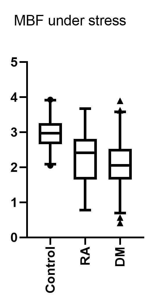
Results: The mean age of patients was 66, with more females in the RA and control group vs the DM group (67% and 69% vs 49% respectively). The total MBF, under adenosine administration, measured in RA patients was higher than DM patients albeit that this did not reach statistical significance ( 2.26 ± -,70 vs 2,05 ± 0,63 mL/min/g, p=0.08 ). When compared to controls, the MBF of RA patients was significantly lower ( 2,94 ± 0,44 vs 2,26 ± 0,70, p < 0.001 ). The coronary flow reserve (CFR) of patients with RA was similar to patients with DM ( 2.20 ± 0,69 vs 2,21 ± 0,70 mL/min/g, p=0.977). The CFR of the control group was significantly higher than those of the RA patients ( 3,03 ± 0,64 vs 2.20 ± 0,69 mL/min/g, p< 0.000 ) and of the DM patients ( 3,03 ± 0,64 vs 2.21 ± 0,70 mL/min/g, p< 0.000 ). 42% of RA- and 38% of DM patients had coronary microvascular dysfunction, compared to 4% in the control group.
Conclusion: Our results indicate an impaired coronary blood flow in both patients with RA and DM in similar levels. Both patient groups had significantly more coronary microvascular dysfunction.
 Figure (1), MBF under stress in mL/min/g
Disclosures: B. Dijkshoorn, None; R. Knol, None; F. Van Der Zant, None; M. Nurmohamed, AbbVie/Abbott; S. Simsek, None.
Background/Purpose: Patients with rheumatoid arthritis (RA) are at increased risk of cardiovascular disease. This risk is similar to that of diabetes mellitus (DM). There have been no studies comparing the coronary microvascular dysfunction assessed with coronary flow reserve (CFR) of RA patients to both patients with DM and a control group (=patients without RA, without DM and no cardiovascular event). Our objective was to assess the difference in coronary microvascular dysfunction in patients with RA in comparison to patients with DM and control group.
Methods: From our 13NH3 myocardial PET/CT registry we included all patients that were included from December 2013 until March 2019. A total of 33 patients with RA, 299 patients with DM and 179 control patients (=patients without RA and without DM) were analyzed. Myocardial blood flow was quantified at rest and under stress induced by administrating adenosine. Coronary flow reserve was calculated by dividing MBF under stress by MBF in rest. CFR < 2 was indicative for coronary microvascular dysfunction.
Results: The mean age of patients was 66, with more females in the RA and control group vs the DM group (67% and 69% vs 49% respectively). The total MBF, under adenosine administration, measured in RA patients was higher than DM patients albeit that this did not reach statistical significance ( 2.26 ± -,70 vs 2,05 ± 0,63 mL/min/g, p=0.08 ). When compared to controls, the MBF of RA patients was significantly lower ( 2,94 ± 0,44 vs 2,26 ± 0,70, p < 0.001 ). The coronary flow reserve (CFR) of patients with RA was similar to patients with DM ( 2.20 ± 0,69 vs 2,21 ± 0,70 mL/min/g, p=0.977). The CFR of the control group was significantly higher than those of the RA patients ( 3,03 ± 0,64 vs 2.20 ± 0,69 mL/min/g, p< 0.000 ) and of the DM patients ( 3,03 ± 0,64 vs 2.21 ± 0,70 mL/min/g, p< 0.000 ). 42% of RA- and 38% of DM patients had coronary microvascular dysfunction, compared to 4% in the control group.
Conclusion: Our results indicate an impaired coronary blood flow in both patients with RA and DM in similar levels. Both patient groups had significantly more coronary microvascular dysfunction.
 Figure (1), MBF under stress in mL/min/g
Disclosures: B. Dijkshoorn, None; R. Knol, None; F. Van Der Zant, None; M. Nurmohamed, AbbVie/Abbott; S. Simsek, None.

