Back
Abstract Session
Vasculitis
Session: Abstracts: Vasculitis – Non-ANCA-Associated and Related Disorders I (1615–1620)
1619: Silent Giant Cell Arteritis in Patients with Polymyalgia Rheumatica: Characteristics and Peculiarities
Sunday, November 13, 2022
5:30 PM – 5:40 PM Eastern Time
Location: Ballroom AB
- ED
Eugenio De Miguel, MD, PhD
Hospital Universitario La Paz
Madrid, Spain
Presenting Author(s)
Eugenio De Miguel1, PIERLUIGI MACCHIONI2, Edoardo Conticini3, Corrado Campochiaro4, Rositsa Karalilova5, Sara Monti6, Cristina Ponte7, Giulia Klinowski2, Irene Monjo8, Paolo Paolo Falsetti3, Anastas Batalov9, alessandro tomelleri4 and Alojzija Hocevar10, 1Hospital Universitario La Paz, Madrid, Spain, 2IRCCS-S.Maria Nuova, Reggio Emilia, Italy, 3Department of Medicine, Surgery and Neurosciences, University of Siena, Siena, Italy, 4IRCCS San Raffaele Hospital. Vita-Salute San Raffaele University, Milano, Italy, 5Medical University of Plovdiv, University Hospital Kaspela, Plovdiv, Bulgaria, 6Rheumatology, Fondazione IRCCS Policlinico S. Matteo, University of Pavia, Pavia, Italy, 7Hospital de Santa Maria, Centro Hospitalar Universitario Lisboa Norte EPE, Lisboa, Portugal, 8Hospital Universitario La Paz - IdiPAZ, Madrid, Spain, 9Medical University of Plovdiv, Plovdiv, Bulgaria, 10University Medical Centre Ljubljana, Ljubljana, Slovenia
Background/Purpose: Polymyalgia rheumatica (PMR) and giant cell arteritis (GCA) are closely related diseases. PMR occurs in approximately 50 % of patients with GCA. In a previous pilot study presented at the ACR 2021 meeting, we observed a 22% prevalence of subclinical GCA in patients with PMR without symptoms or signs suggestive of GCA. The aim of our study is to analyze the characteristics of this subclinical GCA using a bigger sample of patients.
Methods: Cross-sectional multicenter international study of the consecutive newly diagnosed patients with PMR who fulfilled the 2012 EULAR/ACR Provisional Classification Criteria for PMR, without symptoms or signs suggestive of GCA (cohort A). All patients underwent ultrasound (US) of both hips and shoulders, as well as of four bilateral arterial territories: temporal (TA), common carotid (CC), subclavian (SC) and axillary arteries (AX). Patients with a positive halo sign were considered to have subclinical GCA. An intima-media thickness (IMT) ≥0.34 mm for frontal and parietal TA, 0.42 mm for common TA, and ≥1 mm for CC, AX and SC was considered a positive result for halo sign. Clinical, demographic and laboratory characteristics of the PMR pure group were compared with the PMR/subclinical GCA patient group. Cohort B included all consecutive patients with the diagnosis of GCA evaluated in the fast-track clinic of the HULP hospital. Cohort A and B were compared in terms of vessel involvement. Three groups of GCA were established by US: a) isolated cranial-GCA in cases of TA involvement; b) isolated LV-GCA in cases of AX, SC, or CC involvement; and c) mixed pattern-GCA in cases of both cranial and LV involvement.
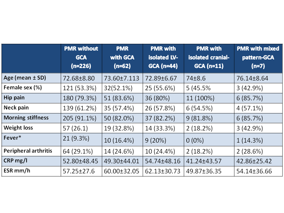
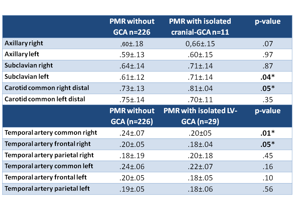
Results: 288 patients were included in cohort A and 97 in cohort B. Table 1 shows the main characteristics of PMR patients (cohort A) with and without subclinical GCA (226 [78.5%] vs 62 [21.5%]), revealing no statistically significant differences between both groups. Only fever showed a significantly higher frequency in subclinical isolated LV-GCA than in cranial-GCA. A trend toward an increased IMT of the LV was observed in patients with PMR and isolated cranial-GCA compared to patients with pure PMR, which was significant in the left SC and right CC (Table 2). However, isolated LV-GCA didn´t show an increase of the IMT in normal TA.
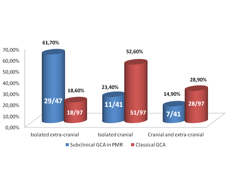
In cohort A, the different subtypes of vessel involvement in patients with PMR were available in 246/288 cases. Data compatible with the diagnosis of GCA was found in 47 cases (19.1%): 11 (23.4%) had isolated cranial-GCA, 29 (61.7%) isolated LV-GCA and 7 (14.9%) mixed pattern-GCA (Figure 1). This was clearly different from our classical cohort of GCA (cohort B), in which the subtype of vessel involvement at presentation was: 51/97 (52.6%) isolated cranial-GCA, 18/97 (18.6%) isolated LV-GCA and 28/97 (28.9%) and mixed pattern-GCA (Figure 1).
Conclusion: A total of 22% of PMR patients without symptoms or signs of GCA have US findings consistent with the diagnosis of GCA. Our data showed a trend for an increased IMT of the large vessels in patients with subclinical GCA and cranial affectation. Subclinical GCA in PMR shows a predilection for extra-cranial artery involvement and has an absolute different ultrasonographic pattern than classical GCA.
 Table 1: Clinical and demographic characteristics of PMR patients with or without GCA
 Table 2: Intima-media thickness measurements in PMR without and with subclinical GCA
 Subtypes of vessel affectation in PMR with subclinical GCA vs Clasical GCA
Disclosures: E. De Miguel, None; P. MACCHIONI, None; E. Conticini, None; C. Campochiaro, None; R. Karalilova, None; S. Monti, None; C. Ponte, None; G. Klinowski, None; I. Monjo, None; P. Paolo Falsetti, None; A. Batalov, Merck/MSD, AbbVie/Abbott, Pfizer, UCB, Novartis, Eli Lilly, Janssen, Roche; a. tomelleri, None; A. Hocevar, None.
Background/Purpose: Polymyalgia rheumatica (PMR) and giant cell arteritis (GCA) are closely related diseases. PMR occurs in approximately 50 % of patients with GCA. In a previous pilot study presented at the ACR 2021 meeting, we observed a 22% prevalence of subclinical GCA in patients with PMR without symptoms or signs suggestive of GCA. The aim of our study is to analyze the characteristics of this subclinical GCA using a bigger sample of patients.
Methods: Cross-sectional multicenter international study of the consecutive newly diagnosed patients with PMR who fulfilled the 2012 EULAR/ACR Provisional Classification Criteria for PMR, without symptoms or signs suggestive of GCA (cohort A). All patients underwent ultrasound (US) of both hips and shoulders, as well as of four bilateral arterial territories: temporal (TA), common carotid (CC), subclavian (SC) and axillary arteries (AX). Patients with a positive halo sign were considered to have subclinical GCA. An intima-media thickness (IMT) ≥0.34 mm for frontal and parietal TA, 0.42 mm for common TA, and ≥1 mm for CC, AX and SC was considered a positive result for halo sign. Clinical, demographic and laboratory characteristics of the PMR pure group were compared with the PMR/subclinical GCA patient group. Cohort B included all consecutive patients with the diagnosis of GCA evaluated in the fast-track clinic of the HULP hospital. Cohort A and B were compared in terms of vessel involvement. Three groups of GCA were established by US: a) isolated cranial-GCA in cases of TA involvement; b) isolated LV-GCA in cases of AX, SC, or CC involvement; and c) mixed pattern-GCA in cases of both cranial and LV involvement.
Results: 288 patients were included in cohort A and 97 in cohort B. Table 1 shows the main characteristics of PMR patients (cohort A) with and without subclinical GCA (226 [78.5%] vs 62 [21.5%]), revealing no statistically significant differences between both groups. Only fever showed a significantly higher frequency in subclinical isolated LV-GCA than in cranial-GCA. A trend toward an increased IMT of the LV was observed in patients with PMR and isolated cranial-GCA compared to patients with pure PMR, which was significant in the left SC and right CC (Table 2). However, isolated LV-GCA didn´t show an increase of the IMT in normal TA.
In cohort A, the different subtypes of vessel involvement in patients with PMR were available in 246/288 cases. Data compatible with the diagnosis of GCA was found in 47 cases (19.1%): 11 (23.4%) had isolated cranial-GCA, 29 (61.7%) isolated LV-GCA and 7 (14.9%) mixed pattern-GCA (Figure 1). This was clearly different from our classical cohort of GCA (cohort B), in which the subtype of vessel involvement at presentation was: 51/97 (52.6%) isolated cranial-GCA, 18/97 (18.6%) isolated LV-GCA and 28/97 (28.9%) and mixed pattern-GCA (Figure 1).
Conclusion: A total of 22% of PMR patients without symptoms or signs of GCA have US findings consistent with the diagnosis of GCA. Our data showed a trend for an increased IMT of the large vessels in patients with subclinical GCA and cranial affectation. Subclinical GCA in PMR shows a predilection for extra-cranial artery involvement and has an absolute different ultrasonographic pattern than classical GCA.
 Table 1: Clinical and demographic characteristics of PMR patients with or without GCA
 Table 2: Intima-media thickness measurements in PMR without and with subclinical GCA
 Subtypes of vessel affectation in PMR with subclinical GCA vs Clasical GCA
Disclosures: E. De Miguel, None; P. MACCHIONI, None; E. Conticini, None; C. Campochiaro, None; R. Karalilova, None; S. Monti, None; C. Ponte, None; G. Klinowski, None; I. Monjo, None; P. Paolo Falsetti, None; A. Batalov, Merck/MSD, AbbVie/Abbott, Pfizer, UCB, Novartis, Eli Lilly, Janssen, Roche; a. tomelleri, None; A. Hocevar, None.

