Back
Poster Session D
Myopathic rheumatic diseases (polymyositis, dermatomyositis, inclusion body myositis)
Session: (1856–1887) Muscle Biology, Myositis and Myopathies Poster II
1879: Analysis of the Association Between the Atrophic Factors Tripartite Motif Containing (TRIM) 63 and Atrogin-1 and the Clinical and Inflammatory Features of Patients with Idiopathic Inflammatory Myopathies
Monday, November 14, 2022
1:00 PM – 3:00 PM Eastern Time
Location: Virtual Poster Hall
- JT
Jiram Torres Ruiz, PhD
INCMNSZ
Mexico, Federal District, Mexico
Abstract Poster Presenter(s)
jiram torres-Ruiz1, Abdiel Absalón-Aguilar2, Juan Alberto Reyes-Islas2, Alfredo Pérez-Fragoso2, Nancy R Mejía-Domínguez3, guillermo Juárez-Vega4, Alejandro Alfaro-Goldaracena5, Beatriz Alcalá-Carmona2, Guillermo Juárez-Vega3, Fabiola Cassiano-Quezada2 and Diana Gómez-Martín1, 1INCMNSZ, Ciudad de México, Mexico, 2Department of Immunology and Rheumatology, Instituto Nacional de Ciencias Médicas y Nutrición Salvador Zubirán, Ciudad de México, Mexico, 3Red de Apoyo a la Investigación, Universidad Nacional Autónoma de México, Ciudad de México, Mexico, 4Red de Apoyo a la Investigación. UNAM, Ciudad de México, Mexico, 5Department of Surgery, Instituto Nacional de Ciencias Médicas y Nutrición Salvador Zubirán, Ciudad de México, Mexico
Background/Purpose: Muscle atrophy is mediated by the ubiquitination of myofilaments by two ubiquitin ligases called Tripartite Motif Containing (TRIM) 63 and Atrogin-1, which are induced by inflammation and interferons (IFN). Muscle atrophy is the main cause of disability in patients with idiopathic inflammatory myopathies (IIM), but notwithstanding its relevance, the expression of TRIM63 and Atrogin-1 and their relationship with atrophy parameters, inflammation and IFN signatures has never been assessed in IIM, which is the aim of the present study.
Methods: For this cross-sectional study, we recruited 37 patients classified as IIM according to the 2017 ACR/EULAR criteria. Patients were clinically evaluated, and we obtained serum, peripheral blood mononuclear cells (PBMCs), and a left deltoid muscle biopsy.
We evaluated the expression of TRIM63 and Atrogin-1 in PBMCs by flow cytometry. The serum levels of the ubiquitin ligases and 17 cytokines and chemokines were addressed by ELISA and a multiplex assay respectively. Atrophy was defined as a muscle fibers' variability index ≥ 250 and an atrophy factor ≥ 150 in men and ≥ 100 in women. In muscle biopsies, we assessed the expression of the ligases, interferon induced gene (ISG)-15, human leukocyte antigen (HLA)-DR, major histocompatibility complex (MHC) II transactivator (CIITA) and MHC-I by immunofluorescence and expressed them as mean fluorescence intensity (MFI). The expression of the ubiquitin ligases, total ubiquitin and the K48 and K63 ubiquitination were analyzed by Western Blot in muscle lysates. As a comparison group, we recruited ten age and sex paired healthy donors. Quantitative variables were compared using the Mann-Whitney U test and correlations were assessed with Spearman Rho.
Results: The median and interquartile range (IQR) of age was 43 years (27-56). Twenty-five patients (67%) were women, and the most frequent diagnosis was dermatomyositis (N=26, 70%). Eight patients (21.6%) had clinically diagnosed muscle atrophy, but muscle biopsies from all patients and none of the healthy donors showed signs of atrophy.
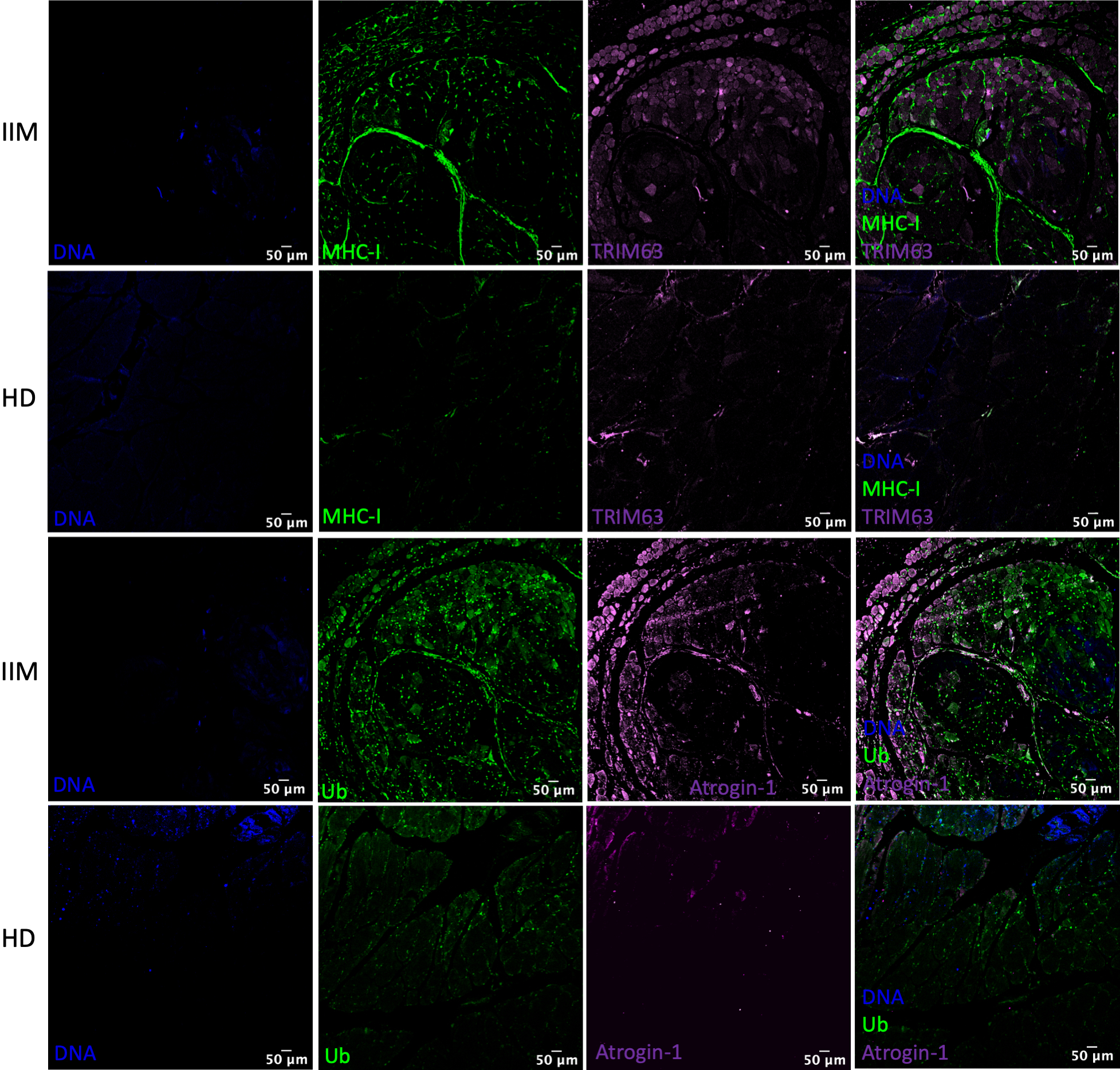
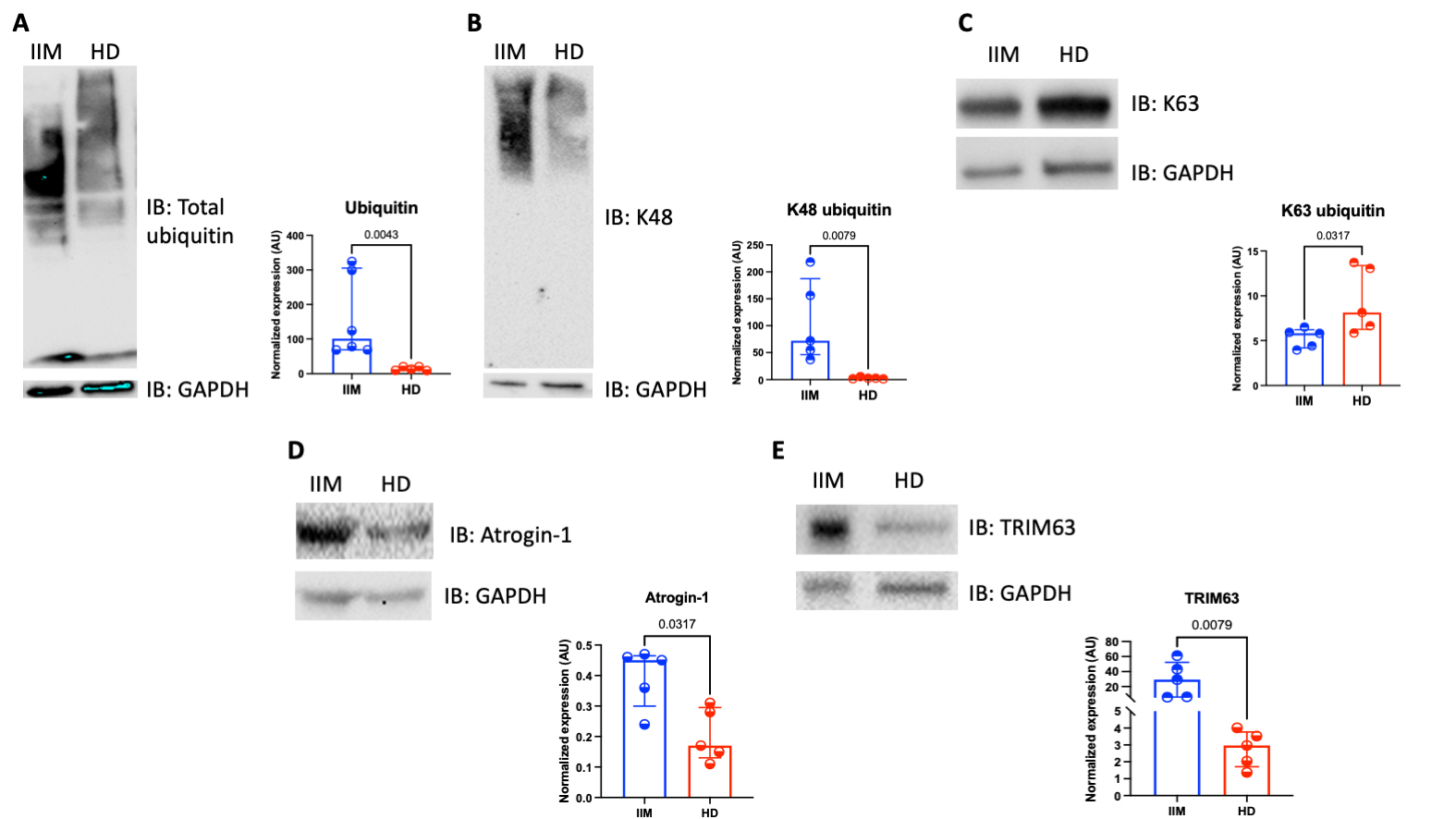
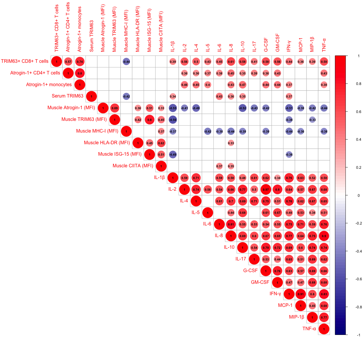
In comparison to healthy donors, IIM patients had a higher muscle expression of TRIM63 and Atrogin-1 (Figure 1). Also, they had a higher normalized expression in arbitrary units of TRIM63 (29.2 (6-52.1) vs 2.9 (1.8-3.7), P=0.007), Atrogin-1 (0.45 (0.3-0.46) vs 0.17 (0.13-0.29), P=0.031), total ubiquitin (100.8 (68.52-305.6) vs 13.3 (9.4-20.4), P=0.004), and K48 ubiquitination (72 (46-187.4) vs 2.7 (1.6-4.3), P=0.007) (Figure 2). In peripheral blood, the percentage of TRIM63+ CD8 T cells correlated with serum cytokines including IL-6 (r=0.45, P=0.004), IFN-γ (r=0.44, P=0.006), and TNF-α (r=0.58, P=0.0001). Serum TRIM63 correlated with IL-6 (r=0.43, P=0.007) and IFN-γ (r=0.37, P=0.023). In muscle tissue, the expression Atrogin-1 and TRIM63 correlated with the interferon markers HLA-DR and ISG-15 (Figure 3). The ubiquitin ligases did not correlate with the atrophy parameters.
Conclusion: In comparison to healthy donors, patients with IIM have a higher expression of TRIM63 and Atrogin-1, which correlates with inflammatory cytokines in peripheral blood and with interferon signatures in muscle biopsies, but not with the muscle parameters of atrophy.
 Figure 1. Confocal microscopy showing a higher expression of TRIM63, Atrogin-1 and ubiquitin in a muscle biopsy from a patient with dermatomyositis in comparison to healthy donor muscle tissue
 Figure 2. Cumulative statistic of the normalized expression of TRIM63, Atrogin-1, total ubiquitin, K48 and K63 ubiquitination in muscle lysates from patients with idiopathic inflammatory myopathies (IIM) and healthy donors (HD).
 Figure 3. Correlation matrix showing the statistically significant correlations between the ubiquitin ligases and the inflammatory parameters of patients with idiopathic inflammatory myopathies
Disclosures: j. torres-Ruiz, None; A. Absalón-Aguilar, None; J. Reyes-Islas, None; A. Pérez-Fragoso, None; N. Mejía-Domínguez, None; g. Juárez-Vega, None; A. Alfaro-Goldaracena, None; B. Alcalá-Carmona, None; G. Juárez-Vega, None; F. Cassiano-Quezada, None; D. Gómez-Martín, None.
Background/Purpose: Muscle atrophy is mediated by the ubiquitination of myofilaments by two ubiquitin ligases called Tripartite Motif Containing (TRIM) 63 and Atrogin-1, which are induced by inflammation and interferons (IFN). Muscle atrophy is the main cause of disability in patients with idiopathic inflammatory myopathies (IIM), but notwithstanding its relevance, the expression of TRIM63 and Atrogin-1 and their relationship with atrophy parameters, inflammation and IFN signatures has never been assessed in IIM, which is the aim of the present study.
Methods: For this cross-sectional study, we recruited 37 patients classified as IIM according to the 2017 ACR/EULAR criteria. Patients were clinically evaluated, and we obtained serum, peripheral blood mononuclear cells (PBMCs), and a left deltoid muscle biopsy.
We evaluated the expression of TRIM63 and Atrogin-1 in PBMCs by flow cytometry. The serum levels of the ubiquitin ligases and 17 cytokines and chemokines were addressed by ELISA and a multiplex assay respectively. Atrophy was defined as a muscle fibers' variability index ≥ 250 and an atrophy factor ≥ 150 in men and ≥ 100 in women. In muscle biopsies, we assessed the expression of the ligases, interferon induced gene (ISG)-15, human leukocyte antigen (HLA)-DR, major histocompatibility complex (MHC) II transactivator (CIITA) and MHC-I by immunofluorescence and expressed them as mean fluorescence intensity (MFI). The expression of the ubiquitin ligases, total ubiquitin and the K48 and K63 ubiquitination were analyzed by Western Blot in muscle lysates. As a comparison group, we recruited ten age and sex paired healthy donors. Quantitative variables were compared using the Mann-Whitney U test and correlations were assessed with Spearman Rho.
Results: The median and interquartile range (IQR) of age was 43 years (27-56). Twenty-five patients (67%) were women, and the most frequent diagnosis was dermatomyositis (N=26, 70%). Eight patients (21.6%) had clinically diagnosed muscle atrophy, but muscle biopsies from all patients and none of the healthy donors showed signs of atrophy.
In comparison to healthy donors, IIM patients had a higher muscle expression of TRIM63 and Atrogin-1 (Figure 1). Also, they had a higher normalized expression in arbitrary units of TRIM63 (29.2 (6-52.1) vs 2.9 (1.8-3.7), P=0.007), Atrogin-1 (0.45 (0.3-0.46) vs 0.17 (0.13-0.29), P=0.031), total ubiquitin (100.8 (68.52-305.6) vs 13.3 (9.4-20.4), P=0.004), and K48 ubiquitination (72 (46-187.4) vs 2.7 (1.6-4.3), P=0.007) (Figure 2). In peripheral blood, the percentage of TRIM63+ CD8 T cells correlated with serum cytokines including IL-6 (r=0.45, P=0.004), IFN-γ (r=0.44, P=0.006), and TNF-α (r=0.58, P=0.0001). Serum TRIM63 correlated with IL-6 (r=0.43, P=0.007) and IFN-γ (r=0.37, P=0.023). In muscle tissue, the expression Atrogin-1 and TRIM63 correlated with the interferon markers HLA-DR and ISG-15 (Figure 3). The ubiquitin ligases did not correlate with the atrophy parameters.
Conclusion: In comparison to healthy donors, patients with IIM have a higher expression of TRIM63 and Atrogin-1, which correlates with inflammatory cytokines in peripheral blood and with interferon signatures in muscle biopsies, but not with the muscle parameters of atrophy.
 Figure 1. Confocal microscopy showing a higher expression of TRIM63, Atrogin-1 and ubiquitin in a muscle biopsy from a patient with dermatomyositis in comparison to healthy donor muscle tissue
 Figure 2. Cumulative statistic of the normalized expression of TRIM63, Atrogin-1, total ubiquitin, K48 and K63 ubiquitination in muscle lysates from patients with idiopathic inflammatory myopathies (IIM) and healthy donors (HD).
  Figure 3. Correlation matrix showing the statistically significant correlations between the ubiquitin ligases and the inflammatory parameters of patients with idiopathic inflammatory myopathies
Disclosures: j. torres-Ruiz, None; A. Absalón-Aguilar, None; J. Reyes-Islas, None; A. Pérez-Fragoso, None; N. Mejía-Domínguez, None; g. Juárez-Vega, None; A. Alfaro-Goldaracena, None; B. Alcalá-Carmona, None; G. Juárez-Vega, None; F. Cassiano-Quezada, None; D. Gómez-Martín, None.

