Back
Poster Session A
Osteoarthritis (OA) and related disorders
Session: (0017–0033) Osteoarthritis and Joint Biology – Basic Science Poster
0018: Longitudinal Development of Cartilage Microbial DNA Profiles Following Oral Microbiome Inoculation
Saturday, November 12, 2022
1:00 PM – 3:00 PM Eastern Time
Location: Virtual Poster Hall
- EP
Emmaline Prinz, BS
University of Oklahoma College of Medicine
Oklahoma City, OK, United States
Abstract Poster Presenter(s)
Emmaline Prinz1, Leoni Schlupp1, Vladislav Izda2, Emily Nguyen1, Christopher Dunn3, Cassandra Sturdy1 and Matlock Jeffries1, 1Oklahoma Medical Research Foundation, Oklahoma City, OK, 2Oklahoma Medical Research Foundation, New York, NY, 3University of Oklahoma Health Sciences Center, Edmond, OK
Background/Purpose: Previous studies have associated microbiome changes with human OA and post-traumatic OA mouse models. Our group has previously published a description of cartilage microbial DNA patterns within both humans and mice, and demonstrated shifts within this microbiome that occur during OA development. In this study, we sought to determine whether and how quickly cartilage microbial DNA patterns develop following inoculation of germ-free animals with a wild-type mouse gut microbiome.
Methods: Germ-free (GF) C57BL6n mice were maintained within the gnotobiotic animal facility at the Oklahoma Medical Research Foundation. 36 female GF animals were inoculated with 1:10 diluted cecal microbiota sourced from adult C57BL6/J mice via oral gavage, then placed in positive pressure cages to pevent contamination. Mice were sacrificed at predetermined intervals following inculation: 4hr (n=6), 24hr (n=6), 48h (n=6), 1wk (n=5), 4wk (n=6), and 8wk (n=6). 7 age-matched uninoculated mice were used as GF controls and 20 non-GF animals from a previous study used as positive controls. Knee articular cartilage was dissected under sterile conditions and ground cryogenically, DNA extracted, and microbial 16S DNA amplified and sequenced. Operational taxonomic units (OTUs) were assigned in QIIME 1.9.1 using the Greengenes 13_8 97% representative reference set. Raw OTU-mapped reads were used to compare inoculated to control animals. Group composition differences were compared by Linear Discriminant Analysis Effect Size (LEfSe, LDA-effect sizes ≥ 2 or ≤ -2 were considered significant) following rarefaction.
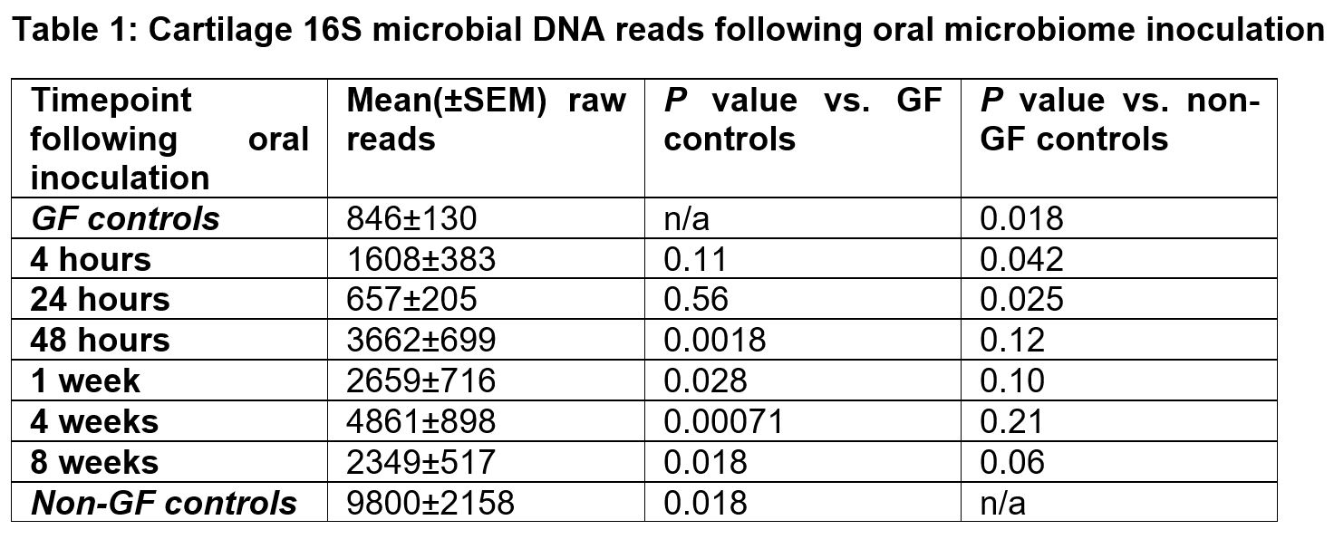
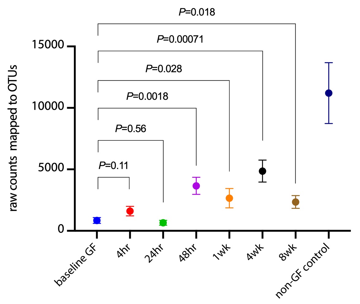
Results: Statistically significant increases in cartilage populations compared to GF mice were noted at the 48h post-inoculation timepoint and later. Similarly, inoculated cartilage populations at the 48h timepoint and later were not statistically significantly different than control non-GF animals (Table 1, Figure 1). 16S cartilage microbiome longitudinal compositional analysis is ongoing.
Conclusion: Cartilage microbial DNA profiles develop rapidly, within 48 hours, following oral microbiome inoculation. Future studies will focus on determining the route of inoculation (presumably, via circulating bloodborne microbes vs. immune trafficking of microbial DNA) and how gut microbial constituents are 'pruned' en route to joints. These findings have important implications for human osteoarthritis (OA) research, where cartilage microbial DNA pattern changes are associated with OA development.
 Table 1: Cartilage 16s microbial DNA reads following oral microbiome inoculation
 Figure 1: Comparison of raw cartilage 16s microbial DNA reads among various oral inoculation times in Germ-free mice
Disclosures: E. Prinz, None; L. Schlupp, None; V. Izda, None; E. Nguyen, None; C. Dunn, None; C. Sturdy, None; M. Jeffries, None.
Background/Purpose: Previous studies have associated microbiome changes with human OA and post-traumatic OA mouse models. Our group has previously published a description of cartilage microbial DNA patterns within both humans and mice, and demonstrated shifts within this microbiome that occur during OA development. In this study, we sought to determine whether and how quickly cartilage microbial DNA patterns develop following inoculation of germ-free animals with a wild-type mouse gut microbiome.
Methods: Germ-free (GF) C57BL6n mice were maintained within the gnotobiotic animal facility at the Oklahoma Medical Research Foundation. 36 female GF animals were inoculated with 1:10 diluted cecal microbiota sourced from adult C57BL6/J mice via oral gavage, then placed in positive pressure cages to pevent contamination. Mice were sacrificed at predetermined intervals following inculation: 4hr (n=6), 24hr (n=6), 48h (n=6), 1wk (n=5), 4wk (n=6), and 8wk (n=6). 7 age-matched uninoculated mice were used as GF controls and 20 non-GF animals from a previous study used as positive controls. Knee articular cartilage was dissected under sterile conditions and ground cryogenically, DNA extracted, and microbial 16S DNA amplified and sequenced. Operational taxonomic units (OTUs) were assigned in QIIME 1.9.1 using the Greengenes 13_8 97% representative reference set. Raw OTU-mapped reads were used to compare inoculated to control animals. Group composition differences were compared by Linear Discriminant Analysis Effect Size (LEfSe, LDA-effect sizes ≥ 2 or ≤ -2 were considered significant) following rarefaction.
Results: Statistically significant increases in cartilage populations compared to GF mice were noted at the 48h post-inoculation timepoint and later. Similarly, inoculated cartilage populations at the 48h timepoint and later were not statistically significantly different than control non-GF animals (Table 1, Figure 1). 16S cartilage microbiome longitudinal compositional analysis is ongoing.
Conclusion: Cartilage microbial DNA profiles develop rapidly, within 48 hours, following oral microbiome inoculation. Future studies will focus on determining the route of inoculation (presumably, via circulating bloodborne microbes vs. immune trafficking of microbial DNA) and how gut microbial constituents are 'pruned' en route to joints. These findings have important implications for human osteoarthritis (OA) research, where cartilage microbial DNA pattern changes are associated with OA development.
 Table 1: Cartilage 16s microbial DNA reads following oral microbiome inoculation
 Figure 1: Comparison of raw cartilage 16s microbial DNA reads among various oral inoculation times in Germ-free mice
Disclosures: E. Prinz, None; L. Schlupp, None; V. Izda, None; E. Nguyen, None; C. Dunn, None; C. Sturdy, None; M. Jeffries, None.

