Back
Poster Session C
Vasculitis
Session: (1543–1578) Vasculitis – Non-ANCA-Associated and Related Disorders Poster II
1557: Microvascular Involvement in Behçet’s Disease: Study of Nailfold Capillaroscopy in Patients from a National Referral Center
Sunday, November 13, 2022
1:00 PM – 3:00 PM Eastern Time
Location: Virtual Poster Hall
- BA
Belen Atienza-Mateo, MD, PhD
Hospital Universitario Marqués de Valdecilla
Santander, Cantabria, Spain
Abstract Poster Presenter(s)
Belén Atienza-Mateo1, Alfonso del Peral-Fanjul2, Veronica Pulito-Cueto3, Diana Prieto-Peña1, Miguel Ángel González-Gay4 and Ricardo Blanco5, 1Research Group on Genetic Epidemiology and Atherosclerosis in Systemic Diseases and in Metabolic Bone Diseases of the Musculoskeletal System, IDIVAL; and Department of Rheumatology, Hospital Universitario Marqués de Valdecilla, Santander, Spain, 2Research Group on Genetic Epidemiology and Atherosclerosis in Systemic Diseases and Metabolic Bone Diseases of the Musculoskeletal System, IDIVAL, Santander, Spain, Spain, 3IDIVAL, Santander, Spain, 4Department of Medicine and Psychiatry, Universidad de Cantabria; Rheumatology Division, Hospital Universitario Marqués de Valdecilla; Research group on genetic epidemiology and atherosclerosis in systemic diseases and in metabolic diseases of the musculoskeletal system, IDIVAL, Santander, Spain. Cardiovascular Pathophysiology and Genomics Research Unit, School of Physiology, Faculty of Health Sciences, University of the Witwatersrand, Johannesburg, South Africa, 5Hospital Universitario Marqués de Valdecilla, IDIVAL, Santander, Spain
Background/Purpose: Ocular and mucocutaneous manifestations of Behçet's disease (BD) are globally recognized and widely studied [Arthritis Rheumatol. 2019 Dec;71(12):2081-2089, Clin Exp Rheumatol. 2020 Sep-Oct;38 Suppl 127(5):69-75]. However, vascular lesions are also frequent in BD, but less explored [Intern Emerg Med. 2019 08;14(5):645-52]. Microvascular involvement may be clinical (as Raynaud's phenomenon) or subclinical. Vascular microcirculation can be studied with Nailfold Capillaroscopy (NFC) and may show capillary abnormalities such as dilated loops, megacapillaries and microhemorrhages. The purpose of our study was to assess the microvascular involvement in patients with BD with and without clinical vascular manifestations.
Methods: Observational study of unselected consecutive patients with BD assessed in a national referral center from March to May 2021. All patients fulfilled the 2014 ICBD criteria. They were evaluated sequentially with a scheduled clinic visit after signing an informed consent. The main periungual capillaries features evaluated were loop diameter, tortuosity, vascular density, microhemorrhages and blood flow. NFC was performed from the second to fifth fingers of both hands using a 500x magnification digital microscope. The following features were considered abnormal: diffuse enlarged capillaries (≥2 capillaries with a diameter of 20-50 µm in at least 2 different fingers), megacapillaries ( >50 µm in diameter), reduced vascular density (< 9 capillaries/mm or avascular areas), diffuse microhemorrhages (≥2 hemorrhages in at least 2 different fingers) and granular/slow flow.
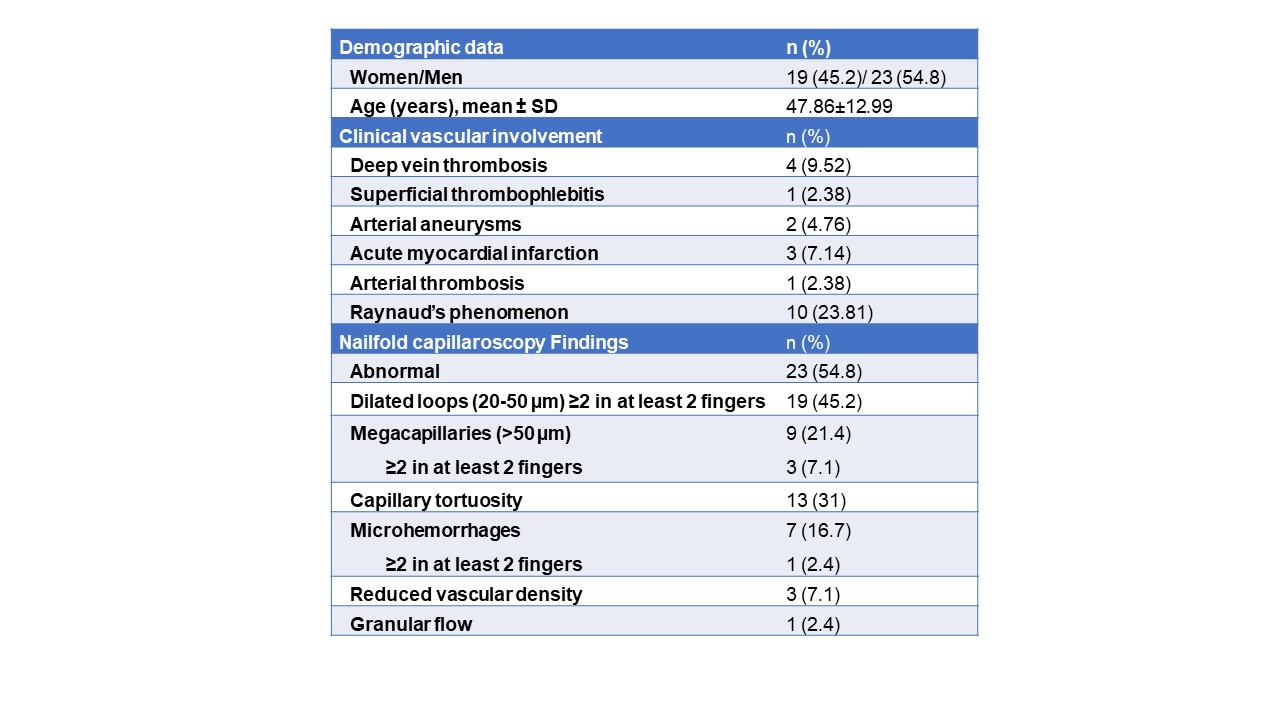
Results: From a cohort of 120 patients with BD, 42 were evaluated during the period of the study (Table). NFC was performed in the 42 cases, being abnormal in 54.8% of them. The most common findings were capillary loop dilation (45.2%), capillary tortuosity (31%), megacapillaries (21.4%) and microhemorrhages (16.7%) (Table and Figure).
Conclusion: Microvascular involvement is not uncommon in BD and can be easily assessed by NFC.
 Table. Clinical vascular and nailfold capillaroscopy data of 42 patients with Behçet’s disease.
.jpg) Figure. Capillaroscopic study images showing, in order, capillary dilations, microhemorrages, and capillary tortuosities.
Figure. Capillaroscopic study images showing, in order, capillary dilations, microhemorrages, and capillary tortuosities.
Disclosures: B. Atienza-Mateo, AbbVie/Abbott, Roche, Pfizer, Celgene, Novartis, Janssen, UCB, Eli Lilly; A. del Peral-Fanjul, None; V. Pulito-Cueto, None; D. Prieto-Peña, UCB, Roche, Pfizer, Amgen, Janssen, AbbVie/Abbott, Novartis, Eli Lilly; M. González-Gay, AbbVie/Abbott, Merck/MSD, Janssen, Roche, AbbVie/Abbott, Roche, Sanofi, Eli Lilly, Celgene, Sobi, Merck/MSD; R. Blanco, Eli Lilly, Pfizer, Roche, Janssen, MSD, AbbVie, Amgen, AstraZeneca, Bristol Myers Squibb, Galapagos, Novartis, Sanofi.
Background/Purpose: Ocular and mucocutaneous manifestations of Behçet's disease (BD) are globally recognized and widely studied [Arthritis Rheumatol. 2019 Dec;71(12):2081-2089, Clin Exp Rheumatol. 2020 Sep-Oct;38 Suppl 127(5):69-75]. However, vascular lesions are also frequent in BD, but less explored [Intern Emerg Med. 2019 08;14(5):645-52]. Microvascular involvement may be clinical (as Raynaud's phenomenon) or subclinical. Vascular microcirculation can be studied with Nailfold Capillaroscopy (NFC) and may show capillary abnormalities such as dilated loops, megacapillaries and microhemorrhages. The purpose of our study was to assess the microvascular involvement in patients with BD with and without clinical vascular manifestations.
Methods: Observational study of unselected consecutive patients with BD assessed in a national referral center from March to May 2021. All patients fulfilled the 2014 ICBD criteria. They were evaluated sequentially with a scheduled clinic visit after signing an informed consent. The main periungual capillaries features evaluated were loop diameter, tortuosity, vascular density, microhemorrhages and blood flow. NFC was performed from the second to fifth fingers of both hands using a 500x magnification digital microscope. The following features were considered abnormal: diffuse enlarged capillaries (≥2 capillaries with a diameter of 20-50 µm in at least 2 different fingers), megacapillaries ( >50 µm in diameter), reduced vascular density (< 9 capillaries/mm or avascular areas), diffuse microhemorrhages (≥2 hemorrhages in at least 2 different fingers) and granular/slow flow.
Results: From a cohort of 120 patients with BD, 42 were evaluated during the period of the study (Table). NFC was performed in the 42 cases, being abnormal in 54.8% of them. The most common findings were capillary loop dilation (45.2%), capillary tortuosity (31%), megacapillaries (21.4%) and microhemorrhages (16.7%) (Table and Figure).
Conclusion: Microvascular involvement is not uncommon in BD and can be easily assessed by NFC.
 Table. Clinical vascular and nailfold capillaroscopy data of 42 patients with Behçet’s disease.
.jpg) Figure. Capillaroscopic study images showing, in order, capillary dilations, microhemorrages, and capillary tortuosities.
Figure. Capillaroscopic study images showing, in order, capillary dilations, microhemorrages, and capillary tortuosities.Disclosures: B. Atienza-Mateo, AbbVie/Abbott, Roche, Pfizer, Celgene, Novartis, Janssen, UCB, Eli Lilly; A. del Peral-Fanjul, None; V. Pulito-Cueto, None; D. Prieto-Peña, UCB, Roche, Pfizer, Amgen, Janssen, AbbVie/Abbott, Novartis, Eli Lilly; M. González-Gay, AbbVie/Abbott, Merck/MSD, Janssen, Roche, AbbVie/Abbott, Roche, Sanofi, Eli Lilly, Celgene, Sobi, Merck/MSD; R. Blanco, Eli Lilly, Pfizer, Roche, Janssen, MSD, AbbVie, Amgen, AstraZeneca, Bristol Myers Squibb, Galapagos, Novartis, Sanofi.

