Back
Ignite Talk
Session: Ignite Session 5C
1065: Biosamples from VEDOSS Patients Show Pathological Signs of SSc: Opportunity for a Biological Diagnosis of Disease
Sunday, November 13, 2022
1:15 PM – 1:20 PM Eastern Time
Location: South Philly Stage

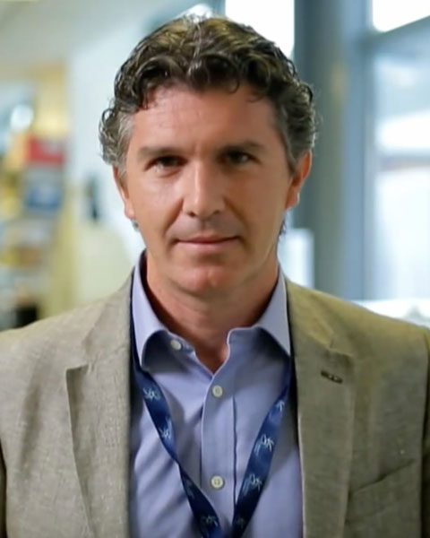
Francesco Del Galdo, MD, PhD
University of Leeds
Leeds, United KingdomDisclosure: Disclosure(s): No relevant disclosure to display
Ignite Speaker(s)
Rebecca Ross1, Emily Clarke1, Will Merchant1, Panji Mulipa1, Natalia Riobo-DelGaldo1 and Francesco Del Galdo2, 1University of Leeds, Leeds, United Kingdom, 2Leeds Institute of Rheumatic and Musculoskeletal Medicine, Faculty of Medicine and Health and NIHR Biomedical Research Centre, University of Leeds, Leeds, United Kingdom
Background/Purpose: The VEDOSS study has recently indicated that more than 50% of patients affected by Raynaud’s phenomenon (RP) and specific SSc anti-nuclear antibodies (ANA) and/or nailfold videocapillaroscopy changes (NVC) (sRP) satisfied ACR/EULAR 2013 criteria within 5 years, indicating a window of opportunity for early detection of SSc. Here we aimed to determine whether sera, skin or dermal fibroblasts cultured from VEDOSS patients showed any biological hallmark of SSc.
Methods: VEDOSS patients were enrolled in the national inception cohort for patients at risk of Scleroderma (Kennedy Cohort, n=64) . Sera were tested for IFN-inducible chemokines (CXCL-9,10 and 11 and CCL2, 8 and 19) and biomarker of extracellular matrix turnover (ELF test) and compared to Healthy control (HC) and SSc sera (n=56, 64, respectively). 3mm skin punch biopsies were taken from the forearms of 13 VEDOSS patients, and 5 healthy controls and 5 SSc patients. Biopsies were analysed for haematoxylin and eosin (H&E), masson trichrome staining (MT) and immunohistological (IHC) staining for CD45. A dermatopathologist blinded to clinical features undertook the analysis. A probabilistic image analysis model was used to detect areas of immunopositivity (CD45) and collagen staining (MT) within the samples, using HeteroGenius Medical Imaging Manager colour analysis. In addition, 7 biopsies (1 ANA-, 6 ANA+), along with 1 healthy control and 3 SSc, were used to explant fibroblasts cultures. mRNA and protein were isolated from primary fibroblasts and processed for RT-qPCR and western blotting analyses.
Results: Sera from sRP patients showed significantly higher IFN-inducible chemokines compared to HC and an intermediate levels compared to SSc [Mean (STDV) HC= 4.69(0.29) vs. VEDOSS= 4.93 (0.39); SSc= 5.30 (0.54)]. Skin biopsies from VEDOSS patients showed evidence of fibrosis, increased collagen bundles within the dermis and increased number of CD45+ cells, as usually seen in SSc, compared to HC. Semiquantitative analysis of histopathological samples indicated a significant correlation between MT and CD45 staining (R=0.8828 P=0.0016). In-vitro, fibroblasts from sRP patients showed an average 5-fold higher collagen mRNA levels and compared to HC fibroblasts, at a similar range to that seen in SSc fibroblasts, confirmed at protein level via western blotting.
Conclusion: Although pilot in nature, this study suggests that patients with no clinical signs of skin fibrosis in their forearms already show biomarker signs of SSc both in their sera, skin biopsies and at dermal fibroblast level. Our data indicate that clinically detectable skin thickening is a late manifestation of SSc pathogenesis and VEDOSS patients offer a biologically active and early window of opportunity for immune and antifibrotic intervention.
Disclosures: R. Ross, None; E. Clarke, None; W. Merchant, None; P. Mulipa, None; N. Riobo-DelGaldo, AstraZeneca; F. Del Galdo, AbbVie/Abbott, AstraZeneca, Boehringer-Ingelheim, Mitsubishi-Tanabe, Capella biosciences, Chemomab LTD, Kymab.

