Back
Poster Session B
Systemic lupus erythematosus (SLE)
Session: (0629–0670) SLE – Etiology and Pathogenesis Poster
0663: Anti-Erythrocyte Antibodies in Childhood Onset Systemic Lupus Erythematosus
Sunday, November 13, 2022
9:00 AM – 10:30 AM Eastern Time
Location: Virtual Poster Hall
- LR
Lauren Robinson, MD
Hospital for Special Surgery
New York, NY, United States
Abstract Poster Presenter(s)
Lauren Robinson1, Zurong Wan2, Preetha Balasubramanian3, Juan Rodriguez Alcazar3, Lynnette Walters4, Jeanine Baisch2, karen onel5, Tracey Wright6, Virginia Pascual3 and Simone Caielli3, 1Hospital for Special Surgery/New York Presbyterian-Weill Cornell, New York, NY, 2Weill Cornell Medicine, New York, NY, 3Weill Cornell Medical College, New York, NY, 4Scottish Rite Hospital for Children, Allen, TX, 5Hospital for Special Surgery, New York, NY, 6UT Southwestern, Plano, TX
Background/Purpose: Systemic Lupus Erythematosus (SLE) is a disease characterized by the presence of auto-antibodies, immune complex deposition, and a robust type 1 interferon signature. It is widely known that SLE patients often have Coombs positivity, suggesting the presence of anti-erythrocyte antibodies. The presence of these antibodies in the absence of hemolytic anemia have long been thought to be clinically insignificant, though an association with a more severe disease phenotype has been reported. Recent work in pediatric SLE has implicated anti-erythrocyte antibodies in disease pathogenesis via opsonization of mitochondria-containing erythrocytes, leading to enhanced erythrophagocytosis and induction of type-1 interferon production by myeloid cells (Caielli et al., 2021). Still, little is known about anti-erythrocyte antibodies in pediatric SLE, including their prevalence, association with disease activity and phenotype, and antigenic target or specificity. The primary objective of this project is to confirm the presence of anti-erythrocyte antibodies in pediatric SLE patients. Additional objectives include characterization of the target antigens and functional properties of these antibodies, as well as to correlate their presence with disease activity and phenotype.
Methods: Serum from 8 pediatric healthy controls and 20 pediatric SLE patients was analyzed for detection of anti-erythrocyte membrane protein antibodies. Erythrocyte membrane protein lysate was generated via the creation of erythrocyte "ghost" membranes and used as a substrate for western blotting. Membranes with erythrocyte membrane proteins were probed with healthy serum or pediatric SLE serum, and the presence of IgG against erythrocyte membrane protein was revealed with anti-human IgG conjugated to HRP.
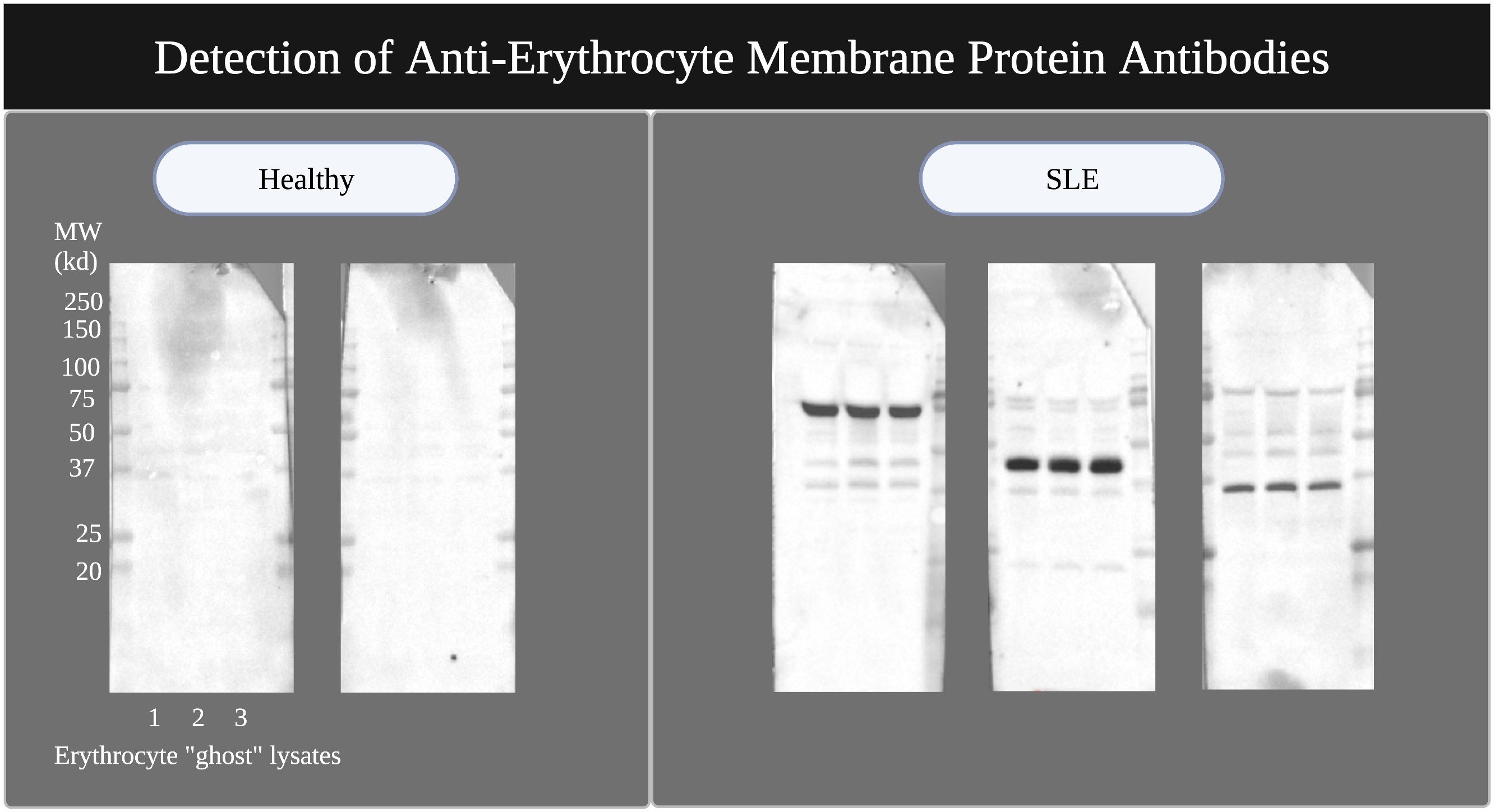
Results: 15 of 20 pediatric SLE serum samples had strongly positive IgG bands to erythrocyte protein antigens on western blot, compared to 0 of 8 healthy pediatric controls. The SLE serum samples showed reactivity against several target antigens of different molecular weights ranging from 25-250kd, and patterns of reactivity varied between samples [Figure 1]. The most prevalent immunoreactive band was at a molecular weight of 75kd (7 of 20 pediatric SLE samples). There was no correlation between the presence of anti-erythrocyte antibodies and hemoglobin or hematocrit.
Conclusion: This study supports the presence of anti-erythrocyte antibodies in a majority of patients with pediatric SLE in the absence of anemia. Future studies will focus on identifying the target erythrocyte proteins and establishing whether a correlation exists between these antibodies and the capacity to opsonize and enhance erythrophagocytosis in vitro. Our ultimate goal is to determine the role these antibodies may play in disease pathogenesis.
 Figure 1: Representative western blots detecting serum antibodies against erythrocyte ghost protein lysate from 3 healthy donors. Left panel shows 2 of 8 blots from pediatric healthy controls. Right panel shows 3 of 20 blots from cSLE patients. All experiments were run in triplicate using ghost membranes from 3 independent donors. Image created with BioRender.
Disclosures: L. Robinson, None; Z. Wan, None; P. Balasubramanian, None; J. Rodriguez Alcazar, None; L. Walters, None; J. Baisch, None; k. onel, None; T. Wright, None; V. Pascual, AstraZeneca, Sanofi, AstraZeneca; S. Caielli, None.
Background/Purpose: Systemic Lupus Erythematosus (SLE) is a disease characterized by the presence of auto-antibodies, immune complex deposition, and a robust type 1 interferon signature. It is widely known that SLE patients often have Coombs positivity, suggesting the presence of anti-erythrocyte antibodies. The presence of these antibodies in the absence of hemolytic anemia have long been thought to be clinically insignificant, though an association with a more severe disease phenotype has been reported. Recent work in pediatric SLE has implicated anti-erythrocyte antibodies in disease pathogenesis via opsonization of mitochondria-containing erythrocytes, leading to enhanced erythrophagocytosis and induction of type-1 interferon production by myeloid cells (Caielli et al., 2021). Still, little is known about anti-erythrocyte antibodies in pediatric SLE, including their prevalence, association with disease activity and phenotype, and antigenic target or specificity. The primary objective of this project is to confirm the presence of anti-erythrocyte antibodies in pediatric SLE patients. Additional objectives include characterization of the target antigens and functional properties of these antibodies, as well as to correlate their presence with disease activity and phenotype.
Methods: Serum from 8 pediatric healthy controls and 20 pediatric SLE patients was analyzed for detection of anti-erythrocyte membrane protein antibodies. Erythrocyte membrane protein lysate was generated via the creation of erythrocyte "ghost" membranes and used as a substrate for western blotting. Membranes with erythrocyte membrane proteins were probed with healthy serum or pediatric SLE serum, and the presence of IgG against erythrocyte membrane protein was revealed with anti-human IgG conjugated to HRP.
Results: 15 of 20 pediatric SLE serum samples had strongly positive IgG bands to erythrocyte protein antigens on western blot, compared to 0 of 8 healthy pediatric controls. The SLE serum samples showed reactivity against several target antigens of different molecular weights ranging from 25-250kd, and patterns of reactivity varied between samples [Figure 1]. The most prevalent immunoreactive band was at a molecular weight of 75kd (7 of 20 pediatric SLE samples). There was no correlation between the presence of anti-erythrocyte antibodies and hemoglobin or hematocrit.
Conclusion: This study supports the presence of anti-erythrocyte antibodies in a majority of patients with pediatric SLE in the absence of anemia. Future studies will focus on identifying the target erythrocyte proteins and establishing whether a correlation exists between these antibodies and the capacity to opsonize and enhance erythrophagocytosis in vitro. Our ultimate goal is to determine the role these antibodies may play in disease pathogenesis.
 Figure 1: Representative western blots detecting serum antibodies against erythrocyte ghost protein lysate from 3 healthy donors. Left panel shows 2 of 8 blots from pediatric healthy controls. Right panel shows 3 of 20 blots from cSLE patients. All experiments were run in triplicate using ghost membranes from 3 independent donors. Image created with BioRender.
Disclosures: L. Robinson, None; Z. Wan, None; P. Balasubramanian, None; J. Rodriguez Alcazar, None; L. Walters, None; J. Baisch, None; k. onel, None; T. Wright, None; V. Pascual, AstraZeneca, Sanofi, AstraZeneca; S. Caielli, None.

