Back
Ignite Talk
Session: Ignite Session 4B
1816: Assessing Urate Deposition and Inflammation in the Vasculature of Gout Patients Using Dual Energy Computed Tomography and Positron Emission Tomography Pre and Post Pegloticase- a Pilot Study
Sunday, November 13, 2022
10:00 AM – 10:05 AM Eastern Time
Location: Center City Stage
- IK
Ira Khanna, MD
Mount Sinai Hospital
New York, NY, United StatesDisclosure: Disclosure information not submitted.
Ignite Speaker(s)
Ira Khanna, Venkatesh Mani, Renata Pyzik, Audrey Kaufman, Wei Wei Chi, Emilia Bagiella, Philip Robson and Yousaf Ali, Icahn School of Medicine at Mount Sinai, New York, NY
Background/Purpose: Gout is the most common inflammatory arthritis, caused by hyperuricemia and subsequent deposition of aggregated monosodium urate (MSU) crystals in both articular and extra-articular regions. A strong association exists between hyperuricemia, gout and risk of cardiovascular disease. CT (DECT) has been shown to detect MSU deposition in vasculature of gout patients with correlation of tophus volume with cardiovascular risk. Vascular Positron Emission Tomography (PET/CT) has been extensively validated to measure arterial vessel wall inflammation. We postulate that uric acid deposits in the vasculature may cause local vascular inflammation. This could be a mechanism involved in theprogression of atherosclerosis and facilitate early onset of adverse cardiovascular events.
Methods: Ten patients with tophaceous gout who were either intolerant or refractory to urate lowering therapy were recruited. Patients were pre-treated with low dose immunomodulator (Azathioprine or Methotrexate) to prevent formation of anti-pegloticase antibodies and then were treated with pegloticase infusions every two weeks for 6 months. DECT and PET/CT were done at baseline and after 6 months of pegloticase to detect vascular MSU deposition (MSU volume) and vessel wall inflammation (Standard uptake value, SUV mean to assess systemic uptake across a vessel segment, SUV max to identify hotspots; Target-to-blood pool ratio, TBR mean and TBR max calculated by dividing the vascular wall SUV with the venous blood pool SUV mean, to correct for blood uptake) respectively. Data were summarized using means and standard deviations. Comparisons of the baseline and follow-up values were conducted for each variable using mixed effect models.
Results: We found a statistically significant decrease in SUV mean (p=0.0003) and SUV max (p=0.0090) across all arteries studied after treatment with pegloticase. There was a trend for decrease in TBR mean and TBR max, which was not statistically significant. In terms of MSU deposition, we found a trend for decrease in MSU volume after treatment with pegloticase, which was not statistically significant.
Conclusion: We observed a statistically significant reduction in vessel wall inflammation (SUV mean and SUV max) along with a trend towards a decrease in MSU deposition (MSU volume) in tophaceous gout patients following treatment with pegloticase. Despite the small sample size of our study, we were able to demonstrate a decrease in both vessel wall inflammation and MSU deposition by aggressively lowering uric acid. These findings suggest a role of MSU in vascular inflammation and demonstrate a concomitant decrease in vessel inflammation as the serum urate level is depleted. It remains to be seen if this correlates to a decrease in adverse cardiovascular outcome.
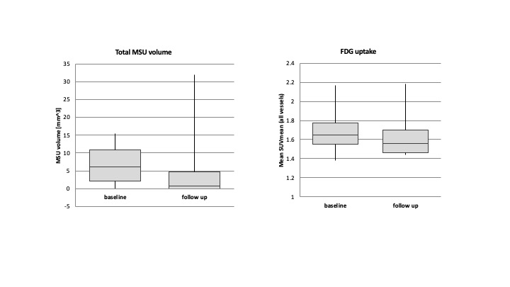
 Figure 1- Bar charts showing decrease in MSU volume after treatment with Pegloticase (p=0.75), and decrease in SUV mean after treatment with Pegloticase (p=0.0003)
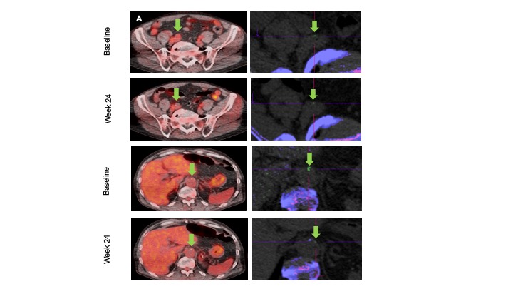
 Figure 2- Representative PET/CT (left) and DECT images (right) from two axial locations in one patient. FDG uptake (orange overlay) shows inflammation in (A) the right iliac artery and (B) abdominal aorta (green arrow). On DECT, MSU deposits (green) are shown in the corresponding vessel walls (cross-hairs), with bone shown purple. In these locations, imaging shows a decrease in FDG uptake and MSU volume following treatment with Pegloticase.
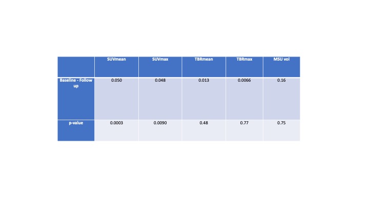
 Table 1- Results of mixed-effects-model analysis. Positive difference (Baseline-Follow up) represents decrease in value at follow up. We observed a statistically significant decrease in SUVmean (p=0.0003) and SUVmax (p=0.0090) at follow up and a trend towards decrease in TBRmean, TBRmax and MSU volume at follow up (not statistically significant).
Disclosures: I. Khanna, None; V. Mani, None; R. Pyzik, None; A. Kaufman, None; W. Chi, None; E. Bagiella, None; P. Robson, None; Y. Ali, None.
Background/Purpose: Gout is the most common inflammatory arthritis, caused by hyperuricemia and subsequent deposition of aggregated monosodium urate (MSU) crystals in both articular and extra-articular regions. A strong association exists between hyperuricemia, gout and risk of cardiovascular disease. CT (DECT) has been shown to detect MSU deposition in vasculature of gout patients with correlation of tophus volume with cardiovascular risk. Vascular Positron Emission Tomography (PET/CT) has been extensively validated to measure arterial vessel wall inflammation. We postulate that uric acid deposits in the vasculature may cause local vascular inflammation. This could be a mechanism involved in theprogression of atherosclerosis and facilitate early onset of adverse cardiovascular events.
Methods: Ten patients with tophaceous gout who were either intolerant or refractory to urate lowering therapy were recruited. Patients were pre-treated with low dose immunomodulator (Azathioprine or Methotrexate) to prevent formation of anti-pegloticase antibodies and then were treated with pegloticase infusions every two weeks for 6 months. DECT and PET/CT were done at baseline and after 6 months of pegloticase to detect vascular MSU deposition (MSU volume) and vessel wall inflammation (Standard uptake value, SUV mean to assess systemic uptake across a vessel segment, SUV max to identify hotspots; Target-to-blood pool ratio, TBR mean and TBR max calculated by dividing the vascular wall SUV with the venous blood pool SUV mean, to correct for blood uptake) respectively. Data were summarized using means and standard deviations. Comparisons of the baseline and follow-up values were conducted for each variable using mixed effect models.
Results: We found a statistically significant decrease in SUV mean (p=0.0003) and SUV max (p=0.0090) across all arteries studied after treatment with pegloticase. There was a trend for decrease in TBR mean and TBR max, which was not statistically significant. In terms of MSU deposition, we found a trend for decrease in MSU volume after treatment with pegloticase, which was not statistically significant.
Conclusion: We observed a statistically significant reduction in vessel wall inflammation (SUV mean and SUV max) along with a trend towards a decrease in MSU deposition (MSU volume) in tophaceous gout patients following treatment with pegloticase. Despite the small sample size of our study, we were able to demonstrate a decrease in both vessel wall inflammation and MSU deposition by aggressively lowering uric acid. These findings suggest a role of MSU in vascular inflammation and demonstrate a concomitant decrease in vessel inflammation as the serum urate level is depleted. It remains to be seen if this correlates to a decrease in adverse cardiovascular outcome.
 Figure 1- Bar charts showing decrease in MSU volume after treatment with Pegloticase (p=0.75), and decrease in SUV mean after treatment with Pegloticase (p=0.0003)
 Figure 2- Representative PET/CT (left) and DECT images (right) from two axial locations in one patient. FDG uptake (orange overlay) shows inflammation in (A) the right iliac artery and (B) abdominal aorta (green arrow). On DECT, MSU deposits (green) are shown in the corresponding vessel walls (cross-hairs), with bone shown purple. In these locations, imaging shows a decrease in FDG uptake and MSU volume following treatment with Pegloticase.
 Table 1- Results of mixed-effects-model analysis. Positive difference (Baseline-Follow up) represents decrease in value at follow up. We observed a statistically significant decrease in SUVmean (p=0.0003) and SUVmax (p=0.0090) at follow up and a trend towards decrease in TBRmean, TBRmax and MSU volume at follow up (not statistically significant).
Disclosures: I. Khanna, None; V. Mani, None; R. Pyzik, None; A. Kaufman, None; W. Chi, None; E. Bagiella, None; P. Robson, None; Y. Ali, None.

