Back
Poster Session C
Imaging
Session: (1228–1266) Imaging of Rheumatic Diseases Poster
1258: Development of International Consensus on a Standardized Image Acquisition Protocol for Diagnostic Evaluation of the Sacroiliac Joints by MRI – an ASAS-SPARTAN Collaboration
Sunday, November 13, 2022
1:00 PM – 3:00 PM Eastern Time
Location: Virtual Poster Hall
- WM
Walter P. Maksymowych, MD
University of Alberta
Edmonton, AB, Canada
Abstract Poster Presenter(s)
Robert G Lambert1, Xenofon Baraliakos2, Stephanie Bernard3, John Carrino4, Torsten Diekhoff5, Iris Eshed6, Kay-Geert Hermann7, Nele Herregods8, Jacob Jaremko1, Lennart Jans8, Anne Jurik9, John O'Neill10, Monique Reijnierse11, Michael Tuite12 and Walter P Maksymowych13, 1University of Alberta, Edmonton, AB, Canada, 2Rheumazentrum Ruhrgebiet Herne, Herne, Germany, 3Brooke Army Medical Center, Fort Sam Houston, TX, 4Hospital for Special Surgery, New York, NY, 5Charité – Universitätsmedizin Berlin, Berlin, Germany, 6Sheba Medical Center, Tel Aviv, Israel, 7Charité - Universitätsmedizin Berlin, Berlin, Germany, 8Ghent University Hospital, Ghent, Belgium, 9Aarhus University Hospital, Aarhus, Denmark, 10McMaster University, Hamilton, ON, Canada, 11Leiden University Medical Center, Leiden, Netherlands, 12University of Wisconsin School of Medicine and Public Health, Madison, WI, 13Department of Medicine, University of Alberta, Edmonton, AB, Canada
Background/Purpose: In 2009, ASAS published a 'Definition of active sacroiliitis on MRI for classification of axial spondyloarthritis (axSpA)'. This definition relied on two MRI sequences to make this determination – semicoronal T1 and STIR. Since then, this approach has frequently been used for diagnosis, even though that was never the intent of the definition. In 2015, the European Society of Skeletal Radiology (ESSR) published its recommendations for an SIJ MRI image acquisition protocol (IAP) for diagnostic purposes that required 4 MRI sequences but there is still no IAP that has been widely accepted as a minimum standard worldwide. In 2020, an informal survey of 24 academic sites (12 Europe, 12 North America) confirmed that 24/24 sites performed a minimum of 3 MRI sequences for diagnosis (19 performed 4-8 sequences) because the 2-sequence protocol was considered inadequate.
Objective: To develop the minimum requirements for a standardized IAP for MRI of the sacroiliac joints for diagnostic ascertainment of sacroiliitis.
Methods: All radiologist members of the ASAS and SPARTAN Classification in axSpA (CLASSIC) project, along with one European and one North American rheumatologist with extensive MRI experience in SpA clinical practice and research, were invited to participate in a consensus exercise. A draft IAP was circulated to all participants along with background information and justification for the draft proposal. Feedback on all issues was received by email, tabulated and recirculated. Participants were broadly in favour of the proposal and two months later a teleconference meeting took place and remaining points of contention were resolved. Examples of the proposed IAP performed on new, 10 and 22 years' old MRI scanners were made available for review in DICOM format. Next the revised draft of the IAP was presented at the ASAS annual meeting to the entire membership on 14 January 2022, and voted on.
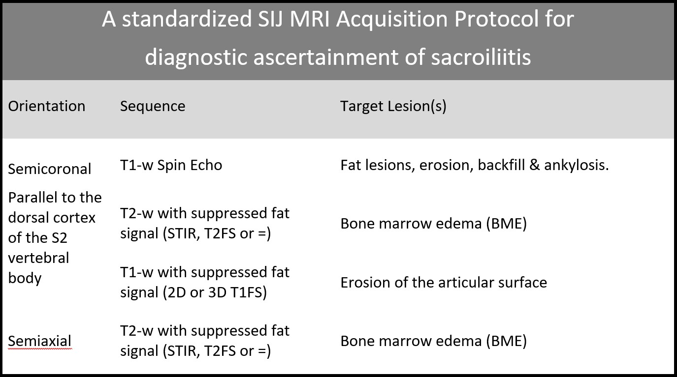
Results: A 4-sequence IAP, 3-semicoronal and 1-semiaxial, is recommended for diagnostic ascertainment of sacroiliitis and its differential diagnoses (Table). It must meet the following requirements: Semicoronal sequences should be parallel to the dorsal cortex of the S2 vertebral body, and include: 1) a sequence sensitive for the detection of active inflammation being t2-weighted with suppression of fat signal; 2) a sequence sensitive for the detection of structural damage in bone and bone marrow with T1-weighting; 3) a sequence that is designed to optimally depict the bone-cartilage interface of the articular surface and be sensitive for detection of bone erosion; and a semiaxial sequence sensitive for inflammation detection. The IAP was approved at the ASAS AGM by a vote of the entire membership with 91% in favour.
Conclusion: A standardized IAP for MRI of the sacroiliac joints for diagnostic ascertainment of sacroiliitis is recommended and should be comprised of 4 sequences, in 2-planes, that will optimally visualize inflammation, structural damage, and the bone-cartilage interface.
 A standardized Sacroiliac Joint MRI Acquisition Protocol for diagnostic ascertainment of sacroiliitis
Disclosures: R. Lambert, Calyx, CARE Arthritis, Image Analysis Group; X. Baraliakos, AbbVie, Lilly, Galapagos, MSD, Novartis, Pfizer, UCB, Bristol-Myers Squibb, Janssen, Roche, Sandoz, Sanofi; S. Bernard, Elsevier; J. Carrino, Pfizer, Regeneron, Globus, Carestream, Image Analysis Group, Image Biopsy Lab; T. Diekhoff, Novartis, Merck/MSD, Canon MS, Eli Lilly; I. Eshed, None; K. Hermann, AbbVie, Merck/MSD, Pfizer, Novartis, BerlinFlame GmbH; N. Herregods, None; J. Jaremko, None; L. Jans, None; A. Jurik, None; J. O'Neill, None; M. Reijnierse, ASAS, International Skeletal Society; M. Tuite, GE HealthCare, Elsevier; W. Maksymowych, AbbVie, Boehringer-Ingelheim, Celgene, Eli Lilly, Galapagos, Janssen, Novartis, Pfizer, UCB, CARE Arthritis Limited.
Background/Purpose: In 2009, ASAS published a 'Definition of active sacroiliitis on MRI for classification of axial spondyloarthritis (axSpA)'. This definition relied on two MRI sequences to make this determination – semicoronal T1 and STIR. Since then, this approach has frequently been used for diagnosis, even though that was never the intent of the definition. In 2015, the European Society of Skeletal Radiology (ESSR) published its recommendations for an SIJ MRI image acquisition protocol (IAP) for diagnostic purposes that required 4 MRI sequences but there is still no IAP that has been widely accepted as a minimum standard worldwide. In 2020, an informal survey of 24 academic sites (12 Europe, 12 North America) confirmed that 24/24 sites performed a minimum of 3 MRI sequences for diagnosis (19 performed 4-8 sequences) because the 2-sequence protocol was considered inadequate.
Objective: To develop the minimum requirements for a standardized IAP for MRI of the sacroiliac joints for diagnostic ascertainment of sacroiliitis.
Methods: All radiologist members of the ASAS and SPARTAN Classification in axSpA (CLASSIC) project, along with one European and one North American rheumatologist with extensive MRI experience in SpA clinical practice and research, were invited to participate in a consensus exercise. A draft IAP was circulated to all participants along with background information and justification for the draft proposal. Feedback on all issues was received by email, tabulated and recirculated. Participants were broadly in favour of the proposal and two months later a teleconference meeting took place and remaining points of contention were resolved. Examples of the proposed IAP performed on new, 10 and 22 years' old MRI scanners were made available for review in DICOM format. Next the revised draft of the IAP was presented at the ASAS annual meeting to the entire membership on 14 January 2022, and voted on.
Results: A 4-sequence IAP, 3-semicoronal and 1-semiaxial, is recommended for diagnostic ascertainment of sacroiliitis and its differential diagnoses (Table). It must meet the following requirements: Semicoronal sequences should be parallel to the dorsal cortex of the S2 vertebral body, and include: 1) a sequence sensitive for the detection of active inflammation being t2-weighted with suppression of fat signal; 2) a sequence sensitive for the detection of structural damage in bone and bone marrow with T1-weighting; 3) a sequence that is designed to optimally depict the bone-cartilage interface of the articular surface and be sensitive for detection of bone erosion; and a semiaxial sequence sensitive for inflammation detection. The IAP was approved at the ASAS AGM by a vote of the entire membership with 91% in favour.
Conclusion: A standardized IAP for MRI of the sacroiliac joints for diagnostic ascertainment of sacroiliitis is recommended and should be comprised of 4 sequences, in 2-planes, that will optimally visualize inflammation, structural damage, and the bone-cartilage interface.
 A standardized Sacroiliac Joint MRI Acquisition Protocol for diagnostic ascertainment of sacroiliitis
Disclosures: R. Lambert, Calyx, CARE Arthritis, Image Analysis Group; X. Baraliakos, AbbVie, Lilly, Galapagos, MSD, Novartis, Pfizer, UCB, Bristol-Myers Squibb, Janssen, Roche, Sandoz, Sanofi; S. Bernard, Elsevier; J. Carrino, Pfizer, Regeneron, Globus, Carestream, Image Analysis Group, Image Biopsy Lab; T. Diekhoff, Novartis, Merck/MSD, Canon MS, Eli Lilly; I. Eshed, None; K. Hermann, AbbVie, Merck/MSD, Pfizer, Novartis, BerlinFlame GmbH; N. Herregods, None; J. Jaremko, None; L. Jans, None; A. Jurik, None; J. O'Neill, None; M. Reijnierse, ASAS, International Skeletal Society; M. Tuite, GE HealthCare, Elsevier; W. Maksymowych, AbbVie, Boehringer-Ingelheim, Celgene, Eli Lilly, Galapagos, Janssen, Novartis, Pfizer, UCB, CARE Arthritis Limited.

