Back
Poster Session A
Vasculitis
Session: (0458–0497) Vasculitis – Non-ANCA-Associated and Related Disorders Poster I: Giant Cell Arteritis
0468: Ultrasound Wall Thickness in the Differential Diagnosis of Atherosclerosis and Large Vessel Giant Cell Arteritis
Saturday, November 12, 2022
1:00 PM – 3:00 PM Eastern Time
Location: Virtual Poster Hall
- ED
Eugenio De Miguel, MD, PhD
Hospital Universitario La Paz
Madrid, Spain
Abstract Poster Presenter(s)
Eugenio De Miguel1, Jose Maria Mostaza2, Carlos Lahoz2, Juan Molina3, Elisa Fernandez-Fernandez1 and Irene Monjo4, 1Hospital Universitario La Paz, Madrid, Spain, 2Hospital Carlos III, Madrid, Spain, 3Hospital General Universitario Gregorio Marañón, Madrid, Spain, 4Hospital Universitario La Paz - IdiPAZ, Madrid, Spain
Background/Purpose: Giant cell arteritis (GCA) is the most common vasculitis in the elderly and large vessel involvement (LV-GCA) occurs in up to 50% of cases. Otherwise, atherosclerosis is frequent in old patients and, as a consequence, ultrasound (US) diagnosis of GCA in these patients may be challenging. The main objective of this study was to determine the validity of US LV-GCA diagnosis using IMT (intima-media thickness)/wall thickness measurements against atherosclerosis.
Methods: We included 47 patients with LV-GCA and 42 controls with atherosclerosis. US examinations of the axillary, subclavian and distal common carotid arteries were systematically performed using a MyLab X8 system (Genoa, Italy) with a 4-15 MHz probe. IMT in atherosclerosis and wall thickness (halo sign) in GCA patients >1mm was accepted as pathological. The maximum IMT in the axillary, subclavian and common carotid arteries (1 cm before the bifurcation) was determined.
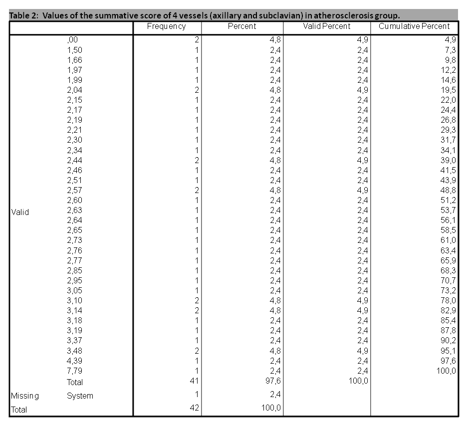
Results: The LV-GCA cohort included 24 females and 23 males with a mean±SD age of 73.8±6.9 years. The atherosclerosis group included 17 males and 25 females with a mean age of 71.8±6.8 years. No significant differences for age (p=0.130) and sex (p=0.395) were found between both cohorts. There were not significant differences in arterial hypertension, diabetes, hyperlipidemia, smoking or obesity between both groups, although the values were numerically higher in the atherosclerosis group. The mean and inter-quartile measures of both cohorts are shown in Table 1. Mean IMT values of all arteries included were significantly higher in patients with LV-GCA than in those with atherosclerosis (Table 1). Among LV-GCA patients, we found axillary involvement in 31 cases (19 bilateral), subclavian in 30 patients (20 bilateral) and common carotid in 28 cases (12 bilateral). In the atherosclerotic cohort we found axillary involvement in 2 cases (1 bilateral), subclavian in 3 cases (none bilateral) and carotid in 14 cases (5 bilateral). We analyzed different scores and a score based on a summative semi-quantitative 0-4 value related with the 20/40/60/80 percentiles measures of axillary and subclavian arteries with a cut-off point ≥ 4 achieved a 95% diagnostic accuracy for GCA with only 2 atherosclerosis patients misdiagnosed (Table 2).
Conclusion: The IMT is higher in LV-GCA than in atherosclerosis and the proposed ultrasound halo score achieves an accuracy > 95% for the differential diagnosis between LV-GCA and atherosclerosis. The axillary and subclavian arteries had a high discriminatory power, while carotid involvement was less confident in the differential diagnosis.
.gif) Table 1: IMT/wall thickness measurements in atherosclerosis and LV-GCA
Table 1: IMT/wall thickness measurements in atherosclerosis and LV-GCA
 Table 2: Values of the summative score of 4 vessels (axillary and subclavian) in atherosclerosis group.
Disclosures: E. De Miguel, None; J. Mostaza, None; C. Lahoz, None; J. Molina, None; E. Fernandez-Fernandez, None; I. Monjo, None.
Background/Purpose: Giant cell arteritis (GCA) is the most common vasculitis in the elderly and large vessel involvement (LV-GCA) occurs in up to 50% of cases. Otherwise, atherosclerosis is frequent in old patients and, as a consequence, ultrasound (US) diagnosis of GCA in these patients may be challenging. The main objective of this study was to determine the validity of US LV-GCA diagnosis using IMT (intima-media thickness)/wall thickness measurements against atherosclerosis.
Methods: We included 47 patients with LV-GCA and 42 controls with atherosclerosis. US examinations of the axillary, subclavian and distal common carotid arteries were systematically performed using a MyLab X8 system (Genoa, Italy) with a 4-15 MHz probe. IMT in atherosclerosis and wall thickness (halo sign) in GCA patients >1mm was accepted as pathological. The maximum IMT in the axillary, subclavian and common carotid arteries (1 cm before the bifurcation) was determined.
Results: The LV-GCA cohort included 24 females and 23 males with a mean±SD age of 73.8±6.9 years. The atherosclerosis group included 17 males and 25 females with a mean age of 71.8±6.8 years. No significant differences for age (p=0.130) and sex (p=0.395) were found between both cohorts. There were not significant differences in arterial hypertension, diabetes, hyperlipidemia, smoking or obesity between both groups, although the values were numerically higher in the atherosclerosis group. The mean and inter-quartile measures of both cohorts are shown in Table 1. Mean IMT values of all arteries included were significantly higher in patients with LV-GCA than in those with atherosclerosis (Table 1). Among LV-GCA patients, we found axillary involvement in 31 cases (19 bilateral), subclavian in 30 patients (20 bilateral) and common carotid in 28 cases (12 bilateral). In the atherosclerotic cohort we found axillary involvement in 2 cases (1 bilateral), subclavian in 3 cases (none bilateral) and carotid in 14 cases (5 bilateral). We analyzed different scores and a score based on a summative semi-quantitative 0-4 value related with the 20/40/60/80 percentiles measures of axillary and subclavian arteries with a cut-off point ≥ 4 achieved a 95% diagnostic accuracy for GCA with only 2 atherosclerosis patients misdiagnosed (Table 2).
Conclusion: The IMT is higher in LV-GCA than in atherosclerosis and the proposed ultrasound halo score achieves an accuracy > 95% for the differential diagnosis between LV-GCA and atherosclerosis. The axillary and subclavian arteries had a high discriminatory power, while carotid involvement was less confident in the differential diagnosis.
.gif) Table 1: IMT/wall thickness measurements in atherosclerosis and LV-GCA
Table 1: IMT/wall thickness measurements in atherosclerosis and LV-GCA Table 2: Values of the summative score of 4 vessels (axillary and subclavian) in atherosclerosis group.
 Disclosures: E. De Miguel, None; J. Mostaza, None; C. Lahoz, None; J. Molina, None; E. Fernandez-Fernandez, None; I. Monjo, None.

