Back
Poster Session A
Spondyloarthritis (SpA) including psoriatic arthritis (PsA)
Session: (0372–0402) Spondyloarthritis Including PsA – Diagnosis, Manifestations, and Outcomes Poster I
0396: Validity of Syndesmophyte Detection on Low Dose Computed Tomography in Patients with Radiographic Axial Spondyloarthritis
Saturday, November 12, 2022
1:00 PM – 3:00 PM Eastern Time
Location: Virtual Poster Hall
- RS
Rosalinde Stal, MSc
Leiden University Medical Center
Leiden, Netherlands
Abstract Poster Presenter(s)
Rosalinde Stal1, Sofia Ramiro1, Xenofon Baraliakos2, Juergen Braun3, Monique Reijnierse1, Rosaline van den Berg1, floris van Gaalen1 and Désirée van der Heijde4, 1Leiden University Medical Center, Leiden, Netherlands, 2Rheumazentrum Ruhrgebiet Herne, Herne, Germany, 3Rheumazentrum Ruhrgebiet, Herne, Germany, 4Department of Rheumatology, Leiden University Medical Center, Leiden, The Netherlands, Leiden, Netherlands
Background/Purpose: Low-dose Computed Tomography (ldCT), assessed with the CT syndesmophyte score (CTSS) in the whole spine, has been shown to detect more progression in the form of new and growing syndesmophytes than the current gold standard, which is conventional radiography (CR) assessed with the modified Stoke Ankylosing Spondylitis Spinal Score (mSASSS). However, the validity of ldCT detected syndesmophytes has not been analyzed. We assessed the convergent validity of ldCT detected syndesmophytes by evaluating whether syndesmophytes on ldCT were also seen on CR at the same vertebral corner (VC) at the same timepoint or two years later.
Methods: Patients from the Sensitive Imaging in Ankylosing Spondylitis cohort underwent ldCT at baseline and CR at baseline and two years. Images were assessed by two (ldCT) or three (CR) readers. While ldCT is able to assess the whole spine and multiple corners per vertebra, in the current study only corners assessed on both modalities by all readers were included to allow for comparison of the two modalities at the VC level. Thus, scores from 12 anterior cervical and 12 anterior lumbar corners were included for each modality. Syndesmophyte presence on ldCT at baseline, regardless of size, was defined per reader and per VC. Syndesmophyte presence on CR, regardless of size, was coded binary as present if seen at baseline, 2 years or both timepoints, per reader per VC. For each reader pair, the number of corners with a syndesmophyte on ldCT at baseline is given, together with the proportion of these corners that also had a syndesmophyte on CR at baseline or 2 years.
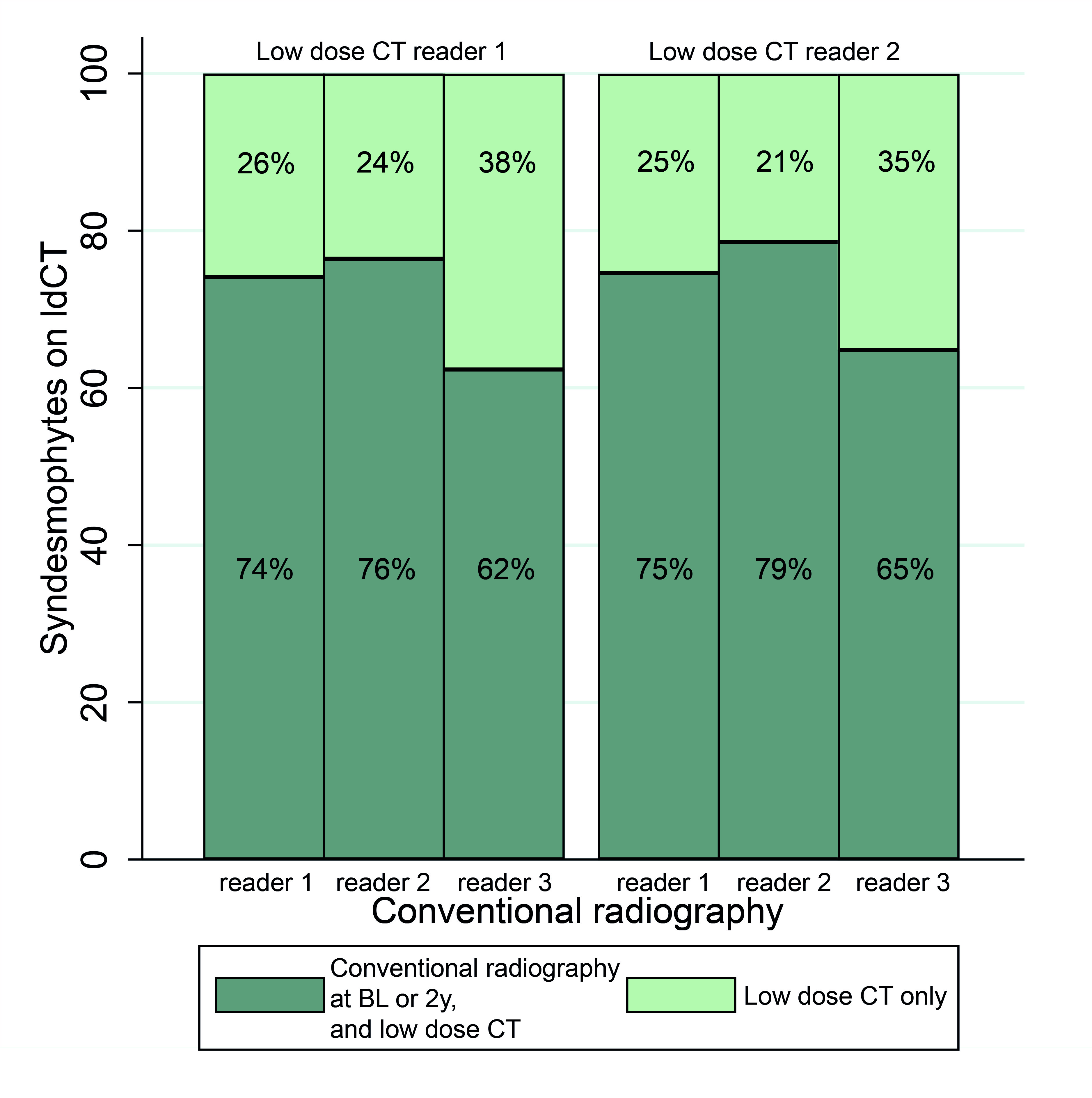
Results: In total 48 patients (85% male, 85% HLA-B27+, mean age 48.3 (SD 9.8) years) had complete imaging and scores, contributing 917 corners per timepoint. At baseline, syndesmophytes were seen on ldCT in 348 (38%, reader 1) and 327 (36%, reader 2) corners out of 917. The figure shows the proportions of corners where a syndesmophyte was also seen on CR at baseline or 2 years per CR reader (62%-79%), and the proportion of corners where a syndesmophyte was only seen on ldCT (21%-38%).
Conclusion: Approximately three quarters of the syndesmophytes visible on ldCT is also visible on the same VC on CR at the same timepoint or two years later. This shows that syndesmophytes on ldCT are also identified as a syndesmophyte on CR on the same VC and adds to the convergent validity of ldCT for the measurement of structural damage in axSpA. Studies with longer follow-up may address whether ldCT syndesmophytes not seen on CR 2 years later can be detected on CR at a later stage.
 Figure: Syndesmophytes visible on low dose Computed Tomography and those also visible on conventional radiography at baseline or 2 years later
The figure displays the corners with a syndesmophyte on low dose CT (reader 1: 348, reader 2: 327) and the proportion of those corners also visible on CR at baseline or 2 years. Results are presented per reader pair. LdCT, low-dose Computed Tomography; BL, baseline; 2y, 2 years.
Disclosures: R. Stal, None; S. Ramiro, AbbVie/Abbott, Eli Lilly, Galapagos, Merck/MSD, Novartis, Pfizer, UCB, Sanofi; X. Baraliakos, AbbVie, Lilly, Galapagos, MSD, Novartis, Pfizer, UCB, Bristol-Myers Squibb, Janssen, Roche, Sandoz, Sanofi; J. Braun, None; M. Reijnierse, ASAS, International Skeletal Society; R. van den Berg, None; f. van Gaalen, Stichting vrienden van Sole Mio, Stichting ASAS, Jacobus stichting, Novartis, UCB, MSD, AbbVie, Bristol Myers Squibb, Eli Lilly; D. van der Heijde, AbbVie, Bayer, BMS, Cyxone, Eisai, Galapagos, Gilead, Glaxo-Smith-Kline, Janssen, Novartis, Pfizer, UCB, Imaging Rheumatology bv, Lilly.
Background/Purpose: Low-dose Computed Tomography (ldCT), assessed with the CT syndesmophyte score (CTSS) in the whole spine, has been shown to detect more progression in the form of new and growing syndesmophytes than the current gold standard, which is conventional radiography (CR) assessed with the modified Stoke Ankylosing Spondylitis Spinal Score (mSASSS). However, the validity of ldCT detected syndesmophytes has not been analyzed. We assessed the convergent validity of ldCT detected syndesmophytes by evaluating whether syndesmophytes on ldCT were also seen on CR at the same vertebral corner (VC) at the same timepoint or two years later.
Methods: Patients from the Sensitive Imaging in Ankylosing Spondylitis cohort underwent ldCT at baseline and CR at baseline and two years. Images were assessed by two (ldCT) or three (CR) readers. While ldCT is able to assess the whole spine and multiple corners per vertebra, in the current study only corners assessed on both modalities by all readers were included to allow for comparison of the two modalities at the VC level. Thus, scores from 12 anterior cervical and 12 anterior lumbar corners were included for each modality. Syndesmophyte presence on ldCT at baseline, regardless of size, was defined per reader and per VC. Syndesmophyte presence on CR, regardless of size, was coded binary as present if seen at baseline, 2 years or both timepoints, per reader per VC. For each reader pair, the number of corners with a syndesmophyte on ldCT at baseline is given, together with the proportion of these corners that also had a syndesmophyte on CR at baseline or 2 years.
Results: In total 48 patients (85% male, 85% HLA-B27+, mean age 48.3 (SD 9.8) years) had complete imaging and scores, contributing 917 corners per timepoint. At baseline, syndesmophytes were seen on ldCT in 348 (38%, reader 1) and 327 (36%, reader 2) corners out of 917. The figure shows the proportions of corners where a syndesmophyte was also seen on CR at baseline or 2 years per CR reader (62%-79%), and the proportion of corners where a syndesmophyte was only seen on ldCT (21%-38%).
Conclusion: Approximately three quarters of the syndesmophytes visible on ldCT is also visible on the same VC on CR at the same timepoint or two years later. This shows that syndesmophytes on ldCT are also identified as a syndesmophyte on CR on the same VC and adds to the convergent validity of ldCT for the measurement of structural damage in axSpA. Studies with longer follow-up may address whether ldCT syndesmophytes not seen on CR 2 years later can be detected on CR at a later stage.
 Figure: Syndesmophytes visible on low dose Computed Tomography and those also visible on conventional radiography at baseline or 2 years later
The figure displays the corners with a syndesmophyte on low dose CT (reader 1: 348, reader 2: 327) and the proportion of those corners also visible on CR at baseline or 2 years. Results are presented per reader pair. LdCT, low-dose Computed Tomography; BL, baseline; 2y, 2 years.
Disclosures: R. Stal, None; S. Ramiro, AbbVie/Abbott, Eli Lilly, Galapagos, Merck/MSD, Novartis, Pfizer, UCB, Sanofi; X. Baraliakos, AbbVie, Lilly, Galapagos, MSD, Novartis, Pfizer, UCB, Bristol-Myers Squibb, Janssen, Roche, Sandoz, Sanofi; J. Braun, None; M. Reijnierse, ASAS, International Skeletal Society; R. van den Berg, None; f. van Gaalen, Stichting vrienden van Sole Mio, Stichting ASAS, Jacobus stichting, Novartis, UCB, MSD, AbbVie, Bristol Myers Squibb, Eli Lilly; D. van der Heijde, AbbVie, Bayer, BMS, Cyxone, Eisai, Galapagos, Gilead, Glaxo-Smith-Kline, Janssen, Novartis, Pfizer, UCB, Imaging Rheumatology bv, Lilly.

