Back
Poster Session A
Myopathic rheumatic diseases (polymyositis, dermatomyositis, inclusion body myositis)
Session: (0150–0180) Muscle Biology, Myositis and Myopathies Poster I
0168: Semiquantitative Thigh Magnetic Resonance Imaging (tMRI) in Determining Skeletal Muscle Outcomes at Baseline and on Follow up in Idiopathic Inflammatory Myopathies (IIMs)
Saturday, November 12, 2022
1:00 PM – 3:00 PM Eastern Time
Location: Virtual Poster Hall
- MG
Mamatha Gorijavolu, MD, MBBS
Jawaharlal Institute of Postgraduate Medical Education and Research
Puducherry, Puducherry, India
Abstract Poster Presenter(s)
Mamatha Gorijavolu1, Devender Bairwa2, Chengappa Kavadichanda3, Sai Kumar Dunga4, Aishwarya Gopal5, Vir Singh Negi6 and Molly Thabah1, 1Jawaharlal Institute of Postgraduate Medical Education and Research, Puducherry, Puducherry, India, 2Jawaharlal Institute of Postgraduate Medical Education and Research (JIPMER), Puducherry, Puducherry, India, 3JIPMER, Pondicherry, Puducherry, India, 4Jawaharlal institute of postgraduate medical education and research, Srikakulam, India, 5Jawaharlal Institute of Postgraduate Medical Education and Research (JIPMER), Pondicherry, Puducherry, India, 6AIIMS Bilaspur, Puducherry, Puducherry, India
Background/Purpose: Semiquantitative scoring of thigh Magnetic Resonance Imaging (tMRI) has shown contradictory results in associating muscle inflammation, damage, and clinically assessed muscle weakness. Moreover, there are no studies assessing the role of tMRI detected muscle damage in determining long-term recovery of muscle strength and endurance.
Objectives: To correlate tMRI scores with concurrently collected manual muscle testing 8 (MMT-8) scores, Two-minute walking Distance(2-MWD) and muscle enzymes. To evaluate the role of baseline tMRI scores in predicting maximum muscle power and 2MWD during follow up.
Methods: This is a retrospective analysis of a single-centre myositis cohort. IIM patients (n=59) who underwent tMRI (STIR and T1 axial and coronal sequences) at the time of diagnosis were included. Baseline demographic, clinical, and serological parameters were noted. MRI images were assessed for intramuscular and fascial edema, muscle atrophy and fatty replacement using a semiquantitative score and the percentage of muscle involvement for each parameter was calculated. On follow up the maximum MMT-8 and 2-MWD for each patient was determined if the two values remained unchanged on 2 separate evaluations at least 3 months apart (n=38). Spearman correlation was done between clinical parameters, muscle enzymes, and tMRI scores. Baseline parameters of patients who achieved and did not achieve maximum MMT8≥74 were compared. Multiple linear regression was performed to assess the tMRI variables predicting MMT-8 and 2-MWD during follow-up.
Results: The median age of the cohort (figure 1) was 38 (30-47) years with a median duration of disease of 4 months (2-12). The Median follow up of the 38 individuals was 31 months (10.00-57.00). Baseline MMT-8 showed strong negative correlation with muscle edema on tMRI (r=-0.755), moderate negative correlation with fascial edema(r=-0.443) and muscle atrophy(r=-0.343). Baseline 2-MWD showed a moderate negative correlation with muscle edema (r=-0.575). Baseline muscle enzymes creatinine kinase (CPK) (r=0.422) and Aspartate transaminase (AST) (r=0.480) showed a moderate positive correlation with tMRI scores for muscle edema. Lactate Dehydrogenase (LDH) (r=0.237) did not significantly correlate with muscle edema. Maximum MMT-8 showed moderate negative correlation with muscle atrophy (r=-0.497) and fatty infiltration(r=-0.531) (Figure2). Follow up 2-MWD showed moderate negative correlation with muscle atrophy(r=-0.353) and fatty infiltration(r=-0.482). Muscle atrophy and fatty infiltration were significantly different between patients who achieved and did not achieve MMT-8 of ≥74 on follow-up (Table 1). Multiple linear regression analysis revealed an adjusted R2 value of 0.655. Muscle atrophy (β=-0.552) and Fatty infiltration (β=-0.418) on baseline tMRI significantly predicted MMT-8 on follow-up independent of other variables.
Conclusion: Muscle loss at baseline in the form of fatty infiltration and atrophy portends poor recovery of muscle power independent of autoantibody status, age and duration.
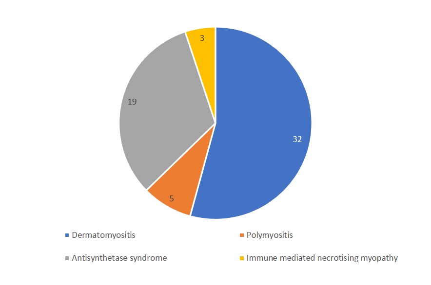
 Figure 1: Distribution of IIM subtypes in the cohort
.jpg) Figure 2: Correlation of baseline and Follow up MMT-8 with tMRI scores
Figure 2: Correlation of baseline and Follow up MMT-8 with tMRI scores
.jpg) Table 1: Comparison between those who achieved near-normal muscle power versus others
Table 1: Comparison between those who achieved near-normal muscle power versus others
Disclosures: M. Gorijavolu, None; D. Bairwa, None; C. Kavadichanda, None; S. Dunga, None; A. Gopal, None; V. Negi, None; M. Thabah, None.
Background/Purpose: Semiquantitative scoring of thigh Magnetic Resonance Imaging (tMRI) has shown contradictory results in associating muscle inflammation, damage, and clinically assessed muscle weakness. Moreover, there are no studies assessing the role of tMRI detected muscle damage in determining long-term recovery of muscle strength and endurance.
Objectives: To correlate tMRI scores with concurrently collected manual muscle testing 8 (MMT-8) scores, Two-minute walking Distance(2-MWD) and muscle enzymes. To evaluate the role of baseline tMRI scores in predicting maximum muscle power and 2MWD during follow up.
Methods: This is a retrospective analysis of a single-centre myositis cohort. IIM patients (n=59) who underwent tMRI (STIR and T1 axial and coronal sequences) at the time of diagnosis were included. Baseline demographic, clinical, and serological parameters were noted. MRI images were assessed for intramuscular and fascial edema, muscle atrophy and fatty replacement using a semiquantitative score and the percentage of muscle involvement for each parameter was calculated. On follow up the maximum MMT-8 and 2-MWD for each patient was determined if the two values remained unchanged on 2 separate evaluations at least 3 months apart (n=38). Spearman correlation was done between clinical parameters, muscle enzymes, and tMRI scores. Baseline parameters of patients who achieved and did not achieve maximum MMT8≥74 were compared. Multiple linear regression was performed to assess the tMRI variables predicting MMT-8 and 2-MWD during follow-up.
Results: The median age of the cohort (figure 1) was 38 (30-47) years with a median duration of disease of 4 months (2-12). The Median follow up of the 38 individuals was 31 months (10.00-57.00). Baseline MMT-8 showed strong negative correlation with muscle edema on tMRI (r=-0.755), moderate negative correlation with fascial edema(r=-0.443) and muscle atrophy(r=-0.343). Baseline 2-MWD showed a moderate negative correlation with muscle edema (r=-0.575). Baseline muscle enzymes creatinine kinase (CPK) (r=0.422) and Aspartate transaminase (AST) (r=0.480) showed a moderate positive correlation with tMRI scores for muscle edema. Lactate Dehydrogenase (LDH) (r=0.237) did not significantly correlate with muscle edema. Maximum MMT-8 showed moderate negative correlation with muscle atrophy (r=-0.497) and fatty infiltration(r=-0.531) (Figure2). Follow up 2-MWD showed moderate negative correlation with muscle atrophy(r=-0.353) and fatty infiltration(r=-0.482). Muscle atrophy and fatty infiltration were significantly different between patients who achieved and did not achieve MMT-8 of ≥74 on follow-up (Table 1). Multiple linear regression analysis revealed an adjusted R2 value of 0.655. Muscle atrophy (β=-0.552) and Fatty infiltration (β=-0.418) on baseline tMRI significantly predicted MMT-8 on follow-up independent of other variables.
Conclusion: Muscle loss at baseline in the form of fatty infiltration and atrophy portends poor recovery of muscle power independent of autoantibody status, age and duration.
 Figure 1: Distribution of IIM subtypes in the cohort
 .jpg) Figure 2: Correlation of baseline and Follow up MMT-8 with tMRI scores
Figure 2: Correlation of baseline and Follow up MMT-8 with tMRI scores.jpg) Table 1: Comparison between those who achieved near-normal muscle power versus others
Table 1: Comparison between those who achieved near-normal muscle power versus others Disclosures: M. Gorijavolu, None; D. Bairwa, None; C. Kavadichanda, None; S. Dunga, None; A. Gopal, None; V. Negi, None; M. Thabah, None.

