Back
Endoscopy Video Forum
Annual Scientific Meeting
Session: Symposium 2C: 10th Annual Endoscopy Video Forum
V4 - Cholangioscopic Recanalization of a Completely Obstructed Post-Transplant Anastomotic Stricture
Monday, October 24, 2022
5:20 PM – 5:30 PM ET
Location: Richardson Ballroom AB

Ghada Mohamed, MBChB
Duke University Medical Center
Durham, NC
Presenting Author - Endoscopy Video Forum(s)
Ghada Mohamed, MBChB1, Andrew Gilman, MD2, Todd H. Baron, MD3
1Duke University Medical Center, Durham, NC; 2University of North Carolina, Chapel Hill, NC; 3University of North Carolina at Chapel Hill, Chapel Hill, NC
Introduction: Liver transplant (LT) is standard of care for patients with end-stage liver disease. However, there is a high rate of biliary complications, especially biliary stenosis. We report a case of a patient presenting with complete biliary obstruction of a duct-to-duct anastomosis successfully managed endoscopically.
Case Description/Methods: A 51-year-old woman underwent orthotopic liver transplantation (OLT) for primary biliary cholangitis. Two months postoperatively she presented with pancreatitis and jaundice. ERCP demonstrated an indwelling surgical biliary “stent” and an anastomotic stricture. The surgical stent was removed, a sphincterotomy was performed, and a 10 mm X 8 cm fully covered stent was placed. Three months later the stent was removed.
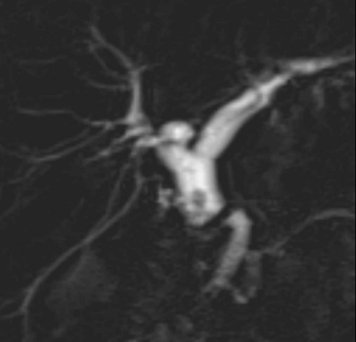
Five months after stent removal the patient presented with fatigue, nausea, abdominal pain and abnormal liver function tests. MRCP showed a severe anastomotic stricture and an upstream stone (Figure 1). ERCP was undertaken. Cholangiography showed a severe anastomotic stricture. Unfortunately, the stricture could not be traversed with 0.025”-0.035” angled hydrophilic guidewires. EUS-guided hepaticogastrostomy was performed and the patient was discharged home.
A follow-up ERCP with cholangioscopy was performed 4 weeks later. At this time contrast would not pass antegrade or retrograde across the anastomosis. Again, guidewires would not traverse the anastomotic stricture with and without cholangioscopic visualization. No lumen could be identified by cholangioscopy. Mini-forceps were used under cholangioscopic direction to recanalize the lumen using a bite-on-bite technique until a lumen was identified. This allowed cholangioscopic guidewire placement for balloon dilation and placement of a 10mm X 10 cm fully covered metal stent.
Discussion: recipients, estimated to occur in up to 30% of patients, with anastomotic strictures being most common.
The mainstay of treatment is endoscopic balloon dilation and biliary stent placement of one covered metal stent or multiple side-by-side plastic stents. However, management is often challenging, with high failure rates. The rate-limiting step for successful therapy is guidewire passage across the stricture. In this case, there was complete obliteration of the lumen by fibrosis such that guidewire passage was not possible.
ERCP with cholangioscopy and mini forceps was used to successfully recanalize the complete anastomotic obstruction and allow for stent placement.

Disclosures:
Ghada Mohamed, MBChB1, Andrew Gilman, MD2, Todd H. Baron, MD3, V4, Cholangioscopic Recanalization of a Completely Obstructed Post-Transplant Anastomotic Stricture, ACG 2022 Annual Scientific Meeting Abstracts. Charlotte, NC: American College of Gastroenterology.
1Duke University Medical Center, Durham, NC; 2University of North Carolina, Chapel Hill, NC; 3University of North Carolina at Chapel Hill, Chapel Hill, NC
Introduction: Liver transplant (LT) is standard of care for patients with end-stage liver disease. However, there is a high rate of biliary complications, especially biliary stenosis. We report a case of a patient presenting with complete biliary obstruction of a duct-to-duct anastomosis successfully managed endoscopically.
Case Description/Methods: A 51-year-old woman underwent orthotopic liver transplantation (OLT) for primary biliary cholangitis. Two months postoperatively she presented with pancreatitis and jaundice. ERCP demonstrated an indwelling surgical biliary “stent” and an anastomotic stricture. The surgical stent was removed, a sphincterotomy was performed, and a 10 mm X 8 cm fully covered stent was placed. Three months later the stent was removed.
Five months after stent removal the patient presented with fatigue, nausea, abdominal pain and abnormal liver function tests. MRCP showed a severe anastomotic stricture and an upstream stone (Figure 1). ERCP was undertaken. Cholangiography showed a severe anastomotic stricture. Unfortunately, the stricture could not be traversed with 0.025”-0.035” angled hydrophilic guidewires. EUS-guided hepaticogastrostomy was performed and the patient was discharged home.
A follow-up ERCP with cholangioscopy was performed 4 weeks later. At this time contrast would not pass antegrade or retrograde across the anastomosis. Again, guidewires would not traverse the anastomotic stricture with and without cholangioscopic visualization. No lumen could be identified by cholangioscopy. Mini-forceps were used under cholangioscopic direction to recanalize the lumen using a bite-on-bite technique until a lumen was identified. This allowed cholangioscopic guidewire placement for balloon dilation and placement of a 10mm X 10 cm fully covered metal stent.
Discussion: recipients, estimated to occur in up to 30% of patients, with anastomotic strictures being most common.
The mainstay of treatment is endoscopic balloon dilation and biliary stent placement of one covered metal stent or multiple side-by-side plastic stents. However, management is often challenging, with high failure rates. The rate-limiting step for successful therapy is guidewire passage across the stricture. In this case, there was complete obliteration of the lumen by fibrosis such that guidewire passage was not possible.
ERCP with cholangioscopy and mini forceps was used to successfully recanalize the complete anastomotic obstruction and allow for stent placement.

Figure: MRCP showing severe anastomotic biliary stricture with an upstream stone.
Disclosures:
Ghada Mohamed indicated no relevant financial relationships.
Andrew Gilman indicated no relevant financial relationships.
Todd Baron: Boston Scientific – Consultant. Cook Endoscopy – Consultant. Olympus America – Consultant. W.L. Gore – Consultant.
Ghada Mohamed, MBChB1, Andrew Gilman, MD2, Todd H. Baron, MD3, V4, Cholangioscopic Recanalization of a Completely Obstructed Post-Transplant Anastomotic Stricture, ACG 2022 Annual Scientific Meeting Abstracts. Charlotte, NC: American College of Gastroenterology.

