Back
Oral Paper Presentation
Annual Scientific Meeting
Session: Plenary Session 4B - Liver
70 - Doppler Ultrasound of the Portal Vein Assessed via Artificial Intelligence With and Without AFP Can Identify Hepatocellular Carcinoma
Wednesday, October 26, 2022
9:45 AM – 10:00 AM ET
Location: Hall C1

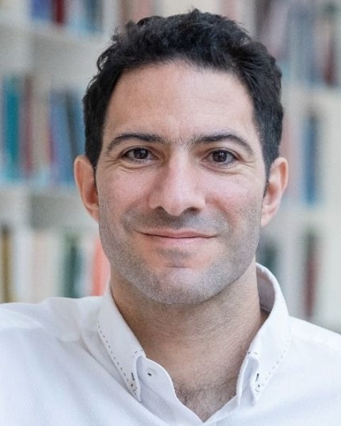
Jose D. Debes, MD, PhD
University of Minnesota
Minneapolis, MN
Presenting Author(s)
Award: ACG Governors Award for Excellence in Clinical Research
Jose D. Debes, MD, PhD1, David Jonason, MD2, Tiancong Cheng, BS1, Ju Sun, PhD1
1University of Minnesota, Minneapolis, MN; 2University of Minnesota Medical Center, Minneapolis, MN
Introduction: Recommendations for hepatocellular carcinoma (HCC) surveillance include assessment of the liver via ultrasound with or without measurement of alpha-fetoprotein (AFP). This approach is operator dependent and difficult to implement in resource-limited settings. We aimed at evaluating if a one-time assessment of the portal vein with sound waves analyzed via artificial intelligence (AI) could aid in HCC detection
Methods: We retrospectively evaluated 142 abdominal ultrasound examinations from our center at University of Minnesota, including 71 liver ultrasounds with HCCs and 71 liver ultrasounds from cirrhotic individuals without HCC. All tumors were confirmed by CT or MRI technology within a period of 6 months of ultrasonography. We included Doppler measurements of the portal vein, hepatic artery and the inferior vena cava. We focused on the analysis of the portal vein due to its large size compared to other vessels and simple approach for detection. As it is standard in AI training procedures, we split the data to 80% as train data and 20% as test data. Due to the limited size of our cohort, we adopted a popular image recognition AI model, VGG-16, pretrained on a large-scale dataset, ImageNet, to extract informative patterns from the measurements. We compared this approach to the measurement of AFP in the same patients
Results: Median age and gender were similar between HCC and control cohorts: 61 years (IQR 55-65) and 59 years (IQR 48-64), as well as 72% and 64% males, respectively. Median size of the HCCs was 2.8cm (IQR 2.3-4.4) which is considered early-stage. Assessment of the portal vein at a cutoff of 20 ng/ml showed an area under the receiving operator curve (AUROC) of 0.85 for HCC detection. A single measurement of the portal vein through our AI algorithm yielded an AUROC of 0.81 for HCC detection. Moreover, a combined AFP and single measurement of PV via AI yielded an impressive AUROC of 0.96 for HCC detection. Assessment of either the hepatic artery or inferior vena cava did not provide a significant AUROC or sensitivity for HCC
Discussion: This is, to our knowledge, the first use of AI for sound analysis in cancer. Our preliminary findings suggest that a single measurement of the portal vein via AI can detect HCC in a similar fashion than AFP and the combination can detect HCC with extremely high accuracy. These findings, if validated in all-size and small HCC, could change our approach to HCC surveillance
Disclosures:
Jose D. Debes, MD, PhD1, David Jonason, MD2, Tiancong Cheng, BS1, Ju Sun, PhD1, 70, Doppler Ultrasound of the Portal Vein Assessed via Artificial Intelligence With and Without AFP Can Identify Hepatocellular Carcinoma, ACG 2022 Annual Scientific Meeting Abstracts. Charlotte, NC: American College of Gastroenterology.
Jose D. Debes, MD, PhD1, David Jonason, MD2, Tiancong Cheng, BS1, Ju Sun, PhD1
1University of Minnesota, Minneapolis, MN; 2University of Minnesota Medical Center, Minneapolis, MN
Introduction: Recommendations for hepatocellular carcinoma (HCC) surveillance include assessment of the liver via ultrasound with or without measurement of alpha-fetoprotein (AFP). This approach is operator dependent and difficult to implement in resource-limited settings. We aimed at evaluating if a one-time assessment of the portal vein with sound waves analyzed via artificial intelligence (AI) could aid in HCC detection
Methods: We retrospectively evaluated 142 abdominal ultrasound examinations from our center at University of Minnesota, including 71 liver ultrasounds with HCCs and 71 liver ultrasounds from cirrhotic individuals without HCC. All tumors were confirmed by CT or MRI technology within a period of 6 months of ultrasonography. We included Doppler measurements of the portal vein, hepatic artery and the inferior vena cava. We focused on the analysis of the portal vein due to its large size compared to other vessels and simple approach for detection. As it is standard in AI training procedures, we split the data to 80% as train data and 20% as test data. Due to the limited size of our cohort, we adopted a popular image recognition AI model, VGG-16, pretrained on a large-scale dataset, ImageNet, to extract informative patterns from the measurements. We compared this approach to the measurement of AFP in the same patients
Results: Median age and gender were similar between HCC and control cohorts: 61 years (IQR 55-65) and 59 years (IQR 48-64), as well as 72% and 64% males, respectively. Median size of the HCCs was 2.8cm (IQR 2.3-4.4) which is considered early-stage. Assessment of the portal vein at a cutoff of 20 ng/ml showed an area under the receiving operator curve (AUROC) of 0.85 for HCC detection. A single measurement of the portal vein through our AI algorithm yielded an AUROC of 0.81 for HCC detection. Moreover, a combined AFP and single measurement of PV via AI yielded an impressive AUROC of 0.96 for HCC detection. Assessment of either the hepatic artery or inferior vena cava did not provide a significant AUROC or sensitivity for HCC
Discussion: This is, to our knowledge, the first use of AI for sound analysis in cancer. Our preliminary findings suggest that a single measurement of the portal vein via AI can detect HCC in a similar fashion than AFP and the combination can detect HCC with extremely high accuracy. These findings, if validated in all-size and small HCC, could change our approach to HCC surveillance
Disclosures:
Jose Debes indicated no relevant financial relationships.
David Jonason indicated no relevant financial relationships.
Tiancong Cheng indicated no relevant financial relationships.
Ju Sun indicated no relevant financial relationships.
Jose D. Debes, MD, PhD1, David Jonason, MD2, Tiancong Cheng, BS1, Ju Sun, PhD1, 70, Doppler Ultrasound of the Portal Vein Assessed via Artificial Intelligence With and Without AFP Can Identify Hepatocellular Carcinoma, ACG 2022 Annual Scientific Meeting Abstracts. Charlotte, NC: American College of Gastroenterology.


