Back
Poster Session A - Sunday Afternoon
A0508 - Isolated Pinworm in Ascites
Sunday, October 23, 2022
5:00 PM – 7:00 PM ET
Location: Crown Ballroom

Priscila Olague, MD
UT HSC - Houston
Houston, TX
Presenting Author(s)
Priscila Olague, MD, Faisal Ali, MD, Violeta Chavez, MD, Tugrul Purnak, MD, Masayuki Nigo, MD
UT HSC - Houston, Houston, TX
Introduction: Extra-intestinal Enterobius vermicularis infections (pinworm) are rare with sites including liver, kidney, spleen, and lung. In females, urinary tract infections and invasion of the genital tract have been described. There have been some case reports describing parasitic infections presenting as eosinophilic ascites in otherwise healthy patients. We present a case in which a cirrhotic patient presented with hepatic encephalopathy (HE) and was found to have a Enterobius vermicularis infection of ascitic fluid.
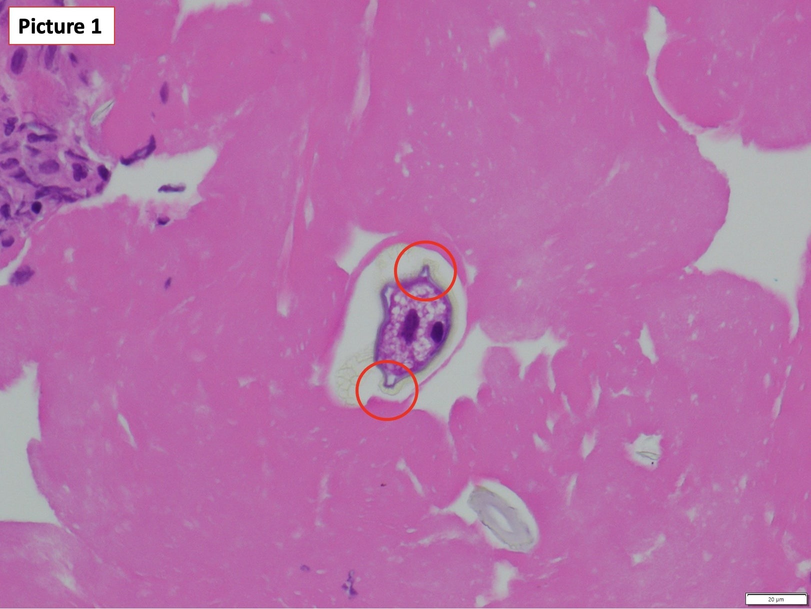
Case Description/Methods: Patient was a 67-year-old female with decompensated cirrhosis from non-alcoholic steatohepatitis (NASH) with ascites, varices, and a history of HE and chronic kidney disease who presented to an outside hospital (OSH) for concerns of HE. The patient had titrated her lactulose from three times to two times daily about one month prior. In addition, diuretics had been held for the past few months due to acute kidney injury (AKI). Abdominal radiography was unremarkable. Laboratory workup at the OSH was unremarkable. A paracentesis was attempted but couldn’t be completed due to the patient's mentation. As she was an established patient, she was transferred to our hospital from the OSH for further management. She initially received lactulose enemas with which her mentation improved. She was eventually placed back on lactulose three times daily, diuretics, and prophylactic antibiotics. A diagnostic paracentesis was performed; fluid studies are shown in Table 1. Cytopathology from paracentesis fluid studies identified Enterobius vermicularis on a cell block section (Figure 1). She developed itching around the umbilical area shortly after her diagnostic paracentesis. She was seen by the infectious diseases team and was prescribed albendazole for a duration of 4 weeks, followed by paracentesis which confirmed eradication.
Discussion: Peritoneal cavity pinworm contamination has been noted presenting as chronic pelvic peritonitis due to enterobius granulomas, however pinworm isolation in ascites is rare. The source in this case is unknown, however, could include translocation from the gut or migration from the genitourinary tract. Treatment with albendazole 400 mg weekly for 4 weeks was effective in eradication of infection.

Disclosures:
Priscila Olague, MD, Faisal Ali, MD, Violeta Chavez, MD, Tugrul Purnak, MD, Masayuki Nigo, MD. A0508 - Isolated Pinworm in Ascites, ACG 2022 Annual Scientific Meeting Abstracts. Charlotte, NC: American College of Gastroenterology.
UT HSC - Houston, Houston, TX
Introduction: Extra-intestinal Enterobius vermicularis infections (pinworm) are rare with sites including liver, kidney, spleen, and lung. In females, urinary tract infections and invasion of the genital tract have been described. There have been some case reports describing parasitic infections presenting as eosinophilic ascites in otherwise healthy patients. We present a case in which a cirrhotic patient presented with hepatic encephalopathy (HE) and was found to have a Enterobius vermicularis infection of ascitic fluid.
Case Description/Methods: Patient was a 67-year-old female with decompensated cirrhosis from non-alcoholic steatohepatitis (NASH) with ascites, varices, and a history of HE and chronic kidney disease who presented to an outside hospital (OSH) for concerns of HE. The patient had titrated her lactulose from three times to two times daily about one month prior. In addition, diuretics had been held for the past few months due to acute kidney injury (AKI). Abdominal radiography was unremarkable. Laboratory workup at the OSH was unremarkable. A paracentesis was attempted but couldn’t be completed due to the patient's mentation. As she was an established patient, she was transferred to our hospital from the OSH for further management. She initially received lactulose enemas with which her mentation improved. She was eventually placed back on lactulose three times daily, diuretics, and prophylactic antibiotics. A diagnostic paracentesis was performed; fluid studies are shown in Table 1. Cytopathology from paracentesis fluid studies identified Enterobius vermicularis on a cell block section (Figure 1). She developed itching around the umbilical area shortly after her diagnostic paracentesis. She was seen by the infectious diseases team and was prescribed albendazole for a duration of 4 weeks, followed by paracentesis which confirmed eradication.
Discussion: Peritoneal cavity pinworm contamination has been noted presenting as chronic pelvic peritonitis due to enterobius granulomas, however pinworm isolation in ascites is rare. The source in this case is unknown, however, could include translocation from the gut or migration from the genitourinary tract. Treatment with albendazole 400 mg weekly for 4 weeks was effective in eradication of infection.

Figure: Picture 1. Pinworm is pictured with characteristic lateral spines in ascites.
Table: Table 1. Ascites fluid studies
Disclosures:
Priscila Olague indicated no relevant financial relationships.
Faisal Ali indicated no relevant financial relationships.
Violeta Chavez indicated no relevant financial relationships.
Tugrul Purnak indicated no relevant financial relationships.
Masayuki Nigo indicated no relevant financial relationships.
Priscila Olague, MD, Faisal Ali, MD, Violeta Chavez, MD, Tugrul Purnak, MD, Masayuki Nigo, MD. A0508 - Isolated Pinworm in Ascites, ACG 2022 Annual Scientific Meeting Abstracts. Charlotte, NC: American College of Gastroenterology.
