Back
Poster Session A - Sunday Afternoon
A0505 - The Imaging Negative Hepatic Lesions: A Rare Case of Infiltrative Hepatocellular Carcinoma
Sunday, October 23, 2022
5:00 PM – 7:00 PM ET
Location: Crown Ballroom

Onyinye Ugonabo, MD
Marshall University Joan C. Edwards School of Medicine
HUNTINGTON, WV
Presenting Author(s)
Onyinye Ugonabo, MD, Joseph Simmons, MD, Saba Altarawneh, MD, Phillip R. Jones, DO, Tejas Joshi, MD, Wesam Frandah, MD
Marshall University Joan C. Edwards School of Medicine, Huntington, WV
Introduction: Hepatocellular Carcinoma (HCC) is the 6th most common cancer and the 4th leading cause of cancer-related death worldwide. The infiltrative subtype is the most difficult to diagnose with imaging because of its inherently ill-defined micronodules involving a segment or entire hepatic parenchyma without a distinguishable mass. Prognosis is poor and estimated at a 5-year survival rate of < 20%. Diagnosis of HCC requires a sensitive imaging test. In situations where imaging is not able to identify a mass, EUS-FNA has been shown to have a higher detection rate.
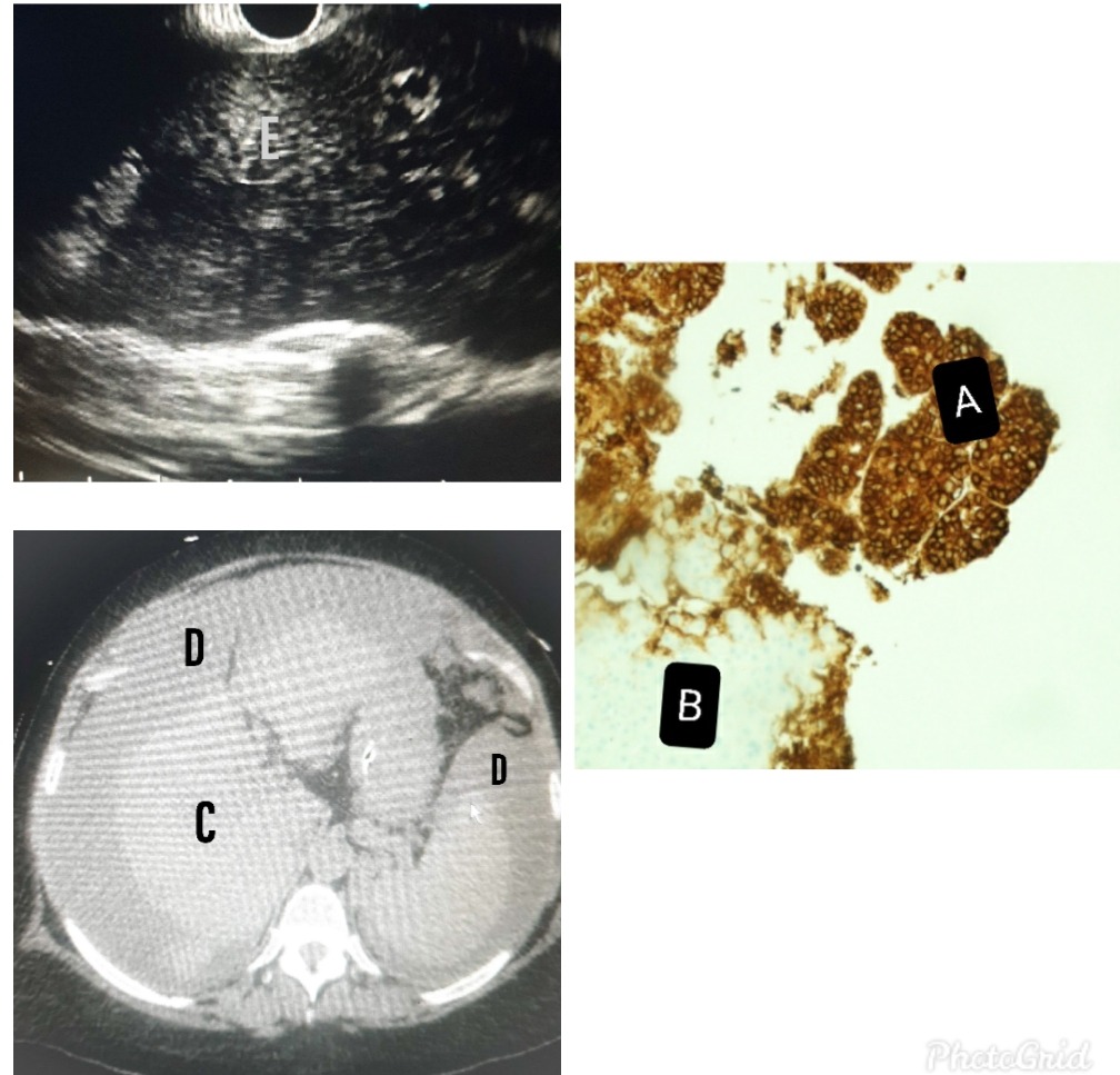
Case Description/Methods: A 61-year-old female with a past medical history of HCV cirrhosis with sustained virology response (SVR), presented with abdominal pain and worsening lower extremity edema. Remarkable examination findings include a distended abdomen and bilateral pitting lower extremity edema. Significant work up showed platelet of 109k/cmm (150-440), AST of 159 unit/l (15-37), total bilirubin of 1.2mg/dl (0.2-1.0), AFP of >20,000ng/ml (0.5-8.0), peritoneal fluid cytology was negative. A right upper quadrant ultrasound, CT, and MRI showed no hepatic mass, fig CD. With markedly elevated AFP and a high suspicion for HCC, the patient had EGD-EUS guided fine-needle aspiration showing multiple infiltrative hepatic lesions, fig E. The biopsy report showed malignant cells positive for AFP with cells reactive for glypican-3 and negative for Hep-Par1 (fig A), supporting the diagnosis of HCC. The patient was referred to oncology and a month later, she died.
Discussion: SVR is associated with decreased risk of HCC, however, with a cirrhotic liver, patients still have an absolute risk of developing HCC. Initial diagnosis of HCC can be obtained non-invasively using abdominal ultrasound, CT, MRI, and EUS-FNA. Abdominal ultrasound is the best imaging modality recommended for HCC surveillance because it is readily available. However, its sensitivity for detecting early HCC is about 47%. AFP cut-off level of > 20ng/ml has a sensitivity of about 60% with low specificity. A level of > 400ng/ml is diagnostic for HCC with a specificity of almost 100%. Incremental changes in AFP are associated with an increased mortality rate. The median survival rate of infiltrative HCC with AFP of > 400 is estimated to be about 5 months. EUS is superior to CT in detecting small hepatic lesions, with a sensitivity of 100% compared to 71% of CT. We recommend that EUS be considered an integral modality while investigating the diagnosis of infiltrative HCC.

Disclosures:
Onyinye Ugonabo, MD, Joseph Simmons, MD, Saba Altarawneh, MD, Phillip R. Jones, DO, Tejas Joshi, MD, Wesam Frandah, MD. A0505 - The Imaging Negative Hepatic Lesions: A Rare Case of Infiltrative Hepatocellular Carcinoma, ACG 2022 Annual Scientific Meeting Abstracts. Charlotte, NC: American College of Gastroenterology.
Marshall University Joan C. Edwards School of Medicine, Huntington, WV
Introduction: Hepatocellular Carcinoma (HCC) is the 6th most common cancer and the 4th leading cause of cancer-related death worldwide. The infiltrative subtype is the most difficult to diagnose with imaging because of its inherently ill-defined micronodules involving a segment or entire hepatic parenchyma without a distinguishable mass. Prognosis is poor and estimated at a 5-year survival rate of < 20%. Diagnosis of HCC requires a sensitive imaging test. In situations where imaging is not able to identify a mass, EUS-FNA has been shown to have a higher detection rate.
Case Description/Methods: A 61-year-old female with a past medical history of HCV cirrhosis with sustained virology response (SVR), presented with abdominal pain and worsening lower extremity edema. Remarkable examination findings include a distended abdomen and bilateral pitting lower extremity edema. Significant work up showed platelet of 109k/cmm (150-440), AST of 159 unit/l (15-37), total bilirubin of 1.2mg/dl (0.2-1.0), AFP of >20,000ng/ml (0.5-8.0), peritoneal fluid cytology was negative. A right upper quadrant ultrasound, CT, and MRI showed no hepatic mass, fig CD. With markedly elevated AFP and a high suspicion for HCC, the patient had EGD-EUS guided fine-needle aspiration showing multiple infiltrative hepatic lesions, fig E. The biopsy report showed malignant cells positive for AFP with cells reactive for glypican-3 and negative for Hep-Par1 (fig A), supporting the diagnosis of HCC. The patient was referred to oncology and a month later, she died.
Discussion: SVR is associated with decreased risk of HCC, however, with a cirrhotic liver, patients still have an absolute risk of developing HCC. Initial diagnosis of HCC can be obtained non-invasively using abdominal ultrasound, CT, MRI, and EUS-FNA. Abdominal ultrasound is the best imaging modality recommended for HCC surveillance because it is readily available. However, its sensitivity for detecting early HCC is about 47%. AFP cut-off level of > 20ng/ml has a sensitivity of about 60% with low specificity. A level of > 400ng/ml is diagnostic for HCC with a specificity of almost 100%. Incremental changes in AFP are associated with an increased mortality rate. The median survival rate of infiltrative HCC with AFP of > 400 is estimated to be about 5 months. EUS is superior to CT in detecting small hepatic lesions, with a sensitivity of 100% compared to 71% of CT. We recommend that EUS be considered an integral modality while investigating the diagnosis of infiltrative HCC.

Figure: Fig A showing Hepatocellular carcinoma, 200x. Glypican-3 immunostain shows strong, diffuse staining in HCC (A). Background non-neoplastic liver is negative (B). Fig C is the cirrhotic liver with no identifiable mass. Fig D shows fluid around the liver and spleen. Fig E is the EUS showing multiple infiltrative hepatic lesions.
Disclosures:
Onyinye Ugonabo indicated no relevant financial relationships.
Joseph Simmons indicated no relevant financial relationships.
Saba Altarawneh indicated no relevant financial relationships.
Phillip Jones indicated no relevant financial relationships.
Tejas Joshi indicated no relevant financial relationships.
Wesam Frandah: Endo gastric solution – Consultant. Olympus – Consultant.
Onyinye Ugonabo, MD, Joseph Simmons, MD, Saba Altarawneh, MD, Phillip R. Jones, DO, Tejas Joshi, MD, Wesam Frandah, MD. A0505 - The Imaging Negative Hepatic Lesions: A Rare Case of Infiltrative Hepatocellular Carcinoma, ACG 2022 Annual Scientific Meeting Abstracts. Charlotte, NC: American College of Gastroenterology.
