Back
Poster Session A - Sunday Afternoon
A0527 - Congenital Riedel’s Lobe of the Liver: A Case Report
Sunday, October 23, 2022
5:00 PM – 7:00 PM ET
Location: Crown Ballroom

Ankoor H. Patel, MD
Rutgers - Robert Wood Johnson Medical School
New Brunswick, NJ
Presenting Author(s)
Ankoor H. Patel, MD1, Rajan N. Amin, MD2, George Abdelsayed, MD1
1Rutgers - Robert Wood Johnson Medical School, New Brunswick, NJ; 2Rutgers University, New Brunswick, NJ
Introduction: Riedel lobe of the liver is a rare anatomical variation with a reported incidence to be between 3.3% and 14.5%. We report a case of a 43-year-old female with an incidental finding of non-palpable Riedel’s lobe.
Case Description/Methods: A 43-year-old female was referred for evaluation of hepatomegaly, which was revealed on MRI and CT scan dating back to 2016. Medical history notable for Irritable Bowel Syndrome (IBS), uterine fibroids, and a history of a tumor removal from her right breast. Patient denies any history of alcohol, illicit drugs, hepatotoxic medications, or pre-existing liver disease. Physical exam was unremarkable and abdominal exam did not reveal any mass or abnormalities.
Routine blood examination, including LFTs and iron studies, was within normal limits. Hepatitis panel (A, B, C), anti-smooth muscle antibody, and LKM-1 IgG antibody was negative. ANA, alpha-1 antitrypsin, and tissue transglutaminase were all negative. The only lab abnormality was an elevated IgG mitochondrial M2 antibody 54.5 (normal less than 20 units).
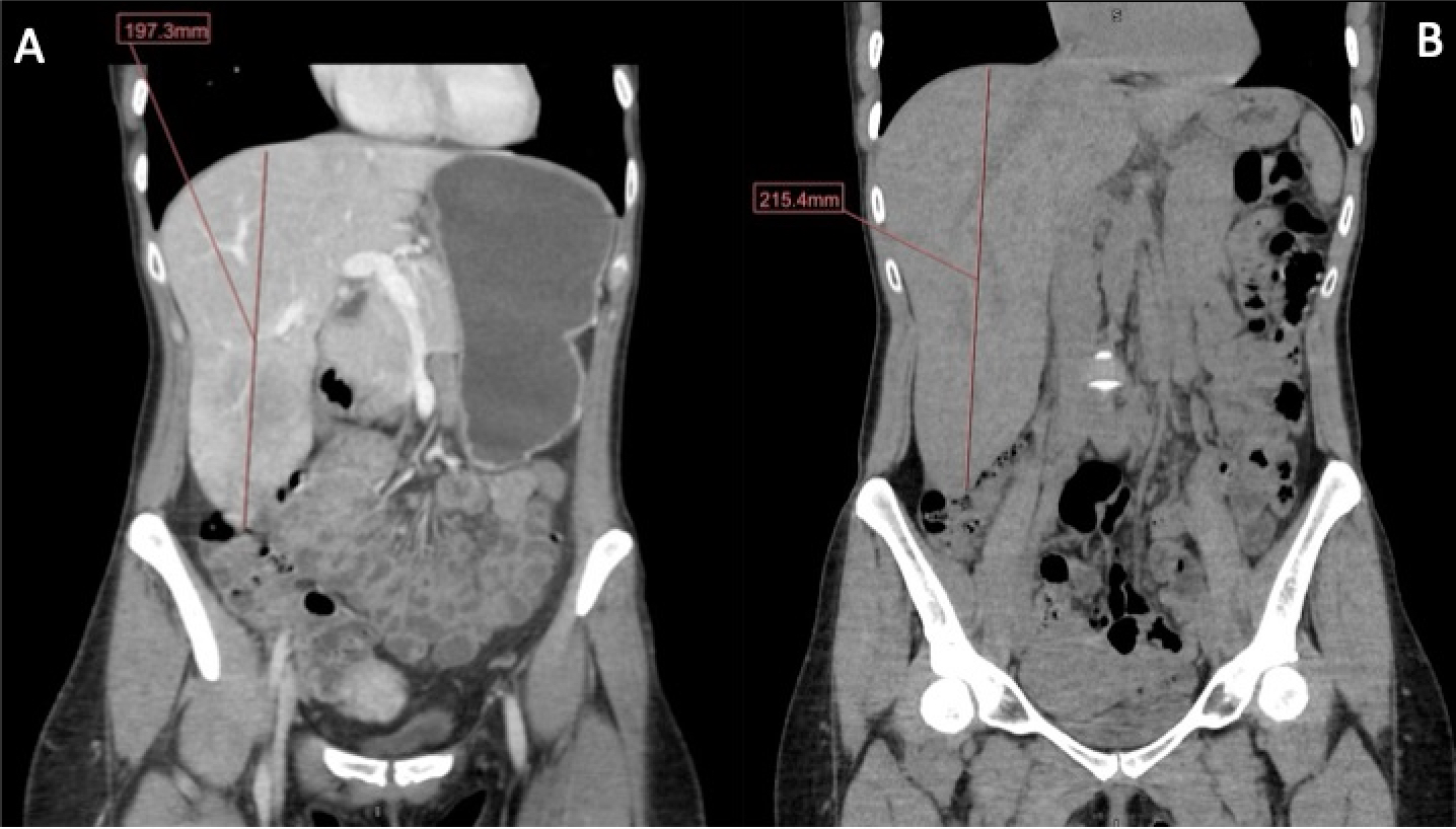
CT abdomen was significant for an enlarged liver with the right lobe extending into the pelvis and ‘not completely included in the study’. MRI abdomen revealed a markedly enlarged liver measuring up to 23.8cm in its craniocaudal dimension with extension into the pelvis with the pancreas deviated to the left, likely secondary to the prominent hepatomegaly. Venous duplex significant for normal directional flow in the portal and hepatic veins with no evidence of portal hypertension. Liver biopsy revealed signs of sinusoidal dilatation nonspecific for veno-occlusive outflow obstruction with no signs of inflammation, steatosis, or fibrosis.
The patient was diagnosed with Riedel’s lobe of the liver. She was discharged from the hospital without treatment with a recommendation to repeat an MRI in 1 year, as torsion is a reported complication of Riedel’s lobe over time. Patient will be recommended to repeat LFTs and anti-mitochondrial antibody to determine progression/significance prior to follow up in 6 months.
Discussion: Riedel’s lobe of the liver is a rare anatomical variant that is often incidentally found on imaging or the presence of hepatomegaly on physical exam. Although patients are usually asymptomatic, its presentation can vary, ranging from nonspecific symptoms to more severe symptoms such as torsion, obstruction, rupture, and bleeding. The range of symptoms highlights its importance in diagnosis and surveillance in this patient population.

Disclosures:
Ankoor H. Patel, MD1, Rajan N. Amin, MD2, George Abdelsayed, MD1. A0527 - Congenital Riedel’s Lobe of the Liver: A Case Report, ACG 2022 Annual Scientific Meeting Abstracts. Charlotte, NC: American College of Gastroenterology.
1Rutgers - Robert Wood Johnson Medical School, New Brunswick, NJ; 2Rutgers University, New Brunswick, NJ
Introduction: Riedel lobe of the liver is a rare anatomical variation with a reported incidence to be between 3.3% and 14.5%. We report a case of a 43-year-old female with an incidental finding of non-palpable Riedel’s lobe.
Case Description/Methods: A 43-year-old female was referred for evaluation of hepatomegaly, which was revealed on MRI and CT scan dating back to 2016. Medical history notable for Irritable Bowel Syndrome (IBS), uterine fibroids, and a history of a tumor removal from her right breast. Patient denies any history of alcohol, illicit drugs, hepatotoxic medications, or pre-existing liver disease. Physical exam was unremarkable and abdominal exam did not reveal any mass or abnormalities.
Routine blood examination, including LFTs and iron studies, was within normal limits. Hepatitis panel (A, B, C), anti-smooth muscle antibody, and LKM-1 IgG antibody was negative. ANA, alpha-1 antitrypsin, and tissue transglutaminase were all negative. The only lab abnormality was an elevated IgG mitochondrial M2 antibody 54.5 (normal less than 20 units).
CT abdomen was significant for an enlarged liver with the right lobe extending into the pelvis and ‘not completely included in the study’. MRI abdomen revealed a markedly enlarged liver measuring up to 23.8cm in its craniocaudal dimension with extension into the pelvis with the pancreas deviated to the left, likely secondary to the prominent hepatomegaly. Venous duplex significant for normal directional flow in the portal and hepatic veins with no evidence of portal hypertension. Liver biopsy revealed signs of sinusoidal dilatation nonspecific for veno-occlusive outflow obstruction with no signs of inflammation, steatosis, or fibrosis.
The patient was diagnosed with Riedel’s lobe of the liver. She was discharged from the hospital without treatment with a recommendation to repeat an MRI in 1 year, as torsion is a reported complication of Riedel’s lobe over time. Patient will be recommended to repeat LFTs and anti-mitochondrial antibody to determine progression/significance prior to follow up in 6 months.
Discussion: Riedel’s lobe of the liver is a rare anatomical variant that is often incidentally found on imaging or the presence of hepatomegaly on physical exam. Although patients are usually asymptomatic, its presentation can vary, ranging from nonspecific symptoms to more severe symptoms such as torsion, obstruction, rupture, and bleeding. The range of symptoms highlights its importance in diagnosis and surveillance in this patient population.

Figure: Figure 1: (A) CT of the abdomen/pelvis from 2016. Liver measuring up to 197.3mm in the sagittal plane. (B) CT of the abdomen/pelvis from 2020. Liver measuring up to 215.4mm in the sagittal plane.
Disclosures:
Ankoor Patel indicated no relevant financial relationships.
Rajan Amin indicated no relevant financial relationships.
George Abdelsayed indicated no relevant financial relationships.
Ankoor H. Patel, MD1, Rajan N. Amin, MD2, George Abdelsayed, MD1. A0527 - Congenital Riedel’s Lobe of the Liver: A Case Report, ACG 2022 Annual Scientific Meeting Abstracts. Charlotte, NC: American College of Gastroenterology.
