Back
Poster Session A - Sunday Afternoon
A0223 - Inlet Patch Polyp Mimicking Esophageal Cancer
Sunday, October 23, 2022
5:00 PM – 7:00 PM ET
Location: Crown Ballroom

Oleksandr Shumeiko, MD
University of Cincinnati
Cincinnati, OH
Presenting Author(s)
Oleksandr Shumeiko, MD1, Serhii Polishchuk, MD2, David Janelidze, MD, PhD2, Yaroslav Dombrovsky, MD3
1University of Cincinnati, Cincinnati, OH; 2GastroCenter Olymed, Kyiv, Kyyiv, Ukraine; 3CSD Medical Laboratory, Kyiv, Kyyiv, Ukraine
Introduction: Heterotopic gastric mucosa (HGM) in the proximal esophagus also known as cervical inlet patch is a prevalent accidental finding during EGD.
Case Description/Methods: 56 y.o. female with no prior PMH presented with a 4-year history of constipation, heartburn, epigastric pain, and weight loss. Symptoms were not controlled with the PPI trial.
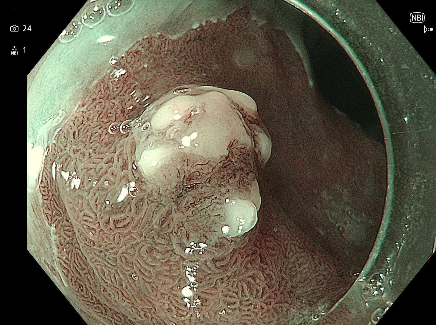
EGD showed 2 areas of gastric mucosa heterotopy in the duodenal bulb, sized up to 5 mm. In the cervical esophagus, an area of HGM sized 15x20 mm with protruding lesion Paris 0-Is sized 5x7mm was visualized. In the narrow-band imaging, the vessel pattern was irregular, with dilated interpupillary loops, and an irregular surface pattern. Malignancy was suspected with biopsies obtained. Pathology showed gastric-type epithelium with active inflammation, erosion, and hyperplastic polyp in HGM. On repeat EGD lesion was completely removed with hot snare polypectomy. Final pathology showed inflammatory (granulation) polyp without dysphasia or neoplastic process. The patient’s dyspeptic symptoms improved on follow-up with double dose PPI.
Discussion: HGM is found in up to 10% of the general population. Some studies consider that HGM is a common benign finding which can be used as a surrogate marker for thorough esophagus examination. Other data suggest that inlet patches can be associated with reflux and dysphagia symptoms, dysplasia, and malignancy in rare cases. Quick insertion and withdrawal of the scope can lead to missed diagnoses of HGM and its complications. HGM detection rate can serve as an EGD quality indicator. Narrow band imaging can be used to improve the HGM detection rate.
Various classifications of surface and vessel patterns are used to identify squamous cell malignancy in the upper esophagus, but they are inapplicable in HGM. Careful examination with histopathological examination of biopsies is recommended for mucosal abnormalities in HGM.

Disclosures:
Oleksandr Shumeiko, MD1, Serhii Polishchuk, MD2, David Janelidze, MD, PhD2, Yaroslav Dombrovsky, MD3. A0223 - Inlet Patch Polyp Mimicking Esophageal Cancer, ACG 2022 Annual Scientific Meeting Abstracts. Charlotte, NC: American College of Gastroenterology.
1University of Cincinnati, Cincinnati, OH; 2GastroCenter Olymed, Kyiv, Kyyiv, Ukraine; 3CSD Medical Laboratory, Kyiv, Kyyiv, Ukraine
Introduction: Heterotopic gastric mucosa (HGM) in the proximal esophagus also known as cervical inlet patch is a prevalent accidental finding during EGD.
Case Description/Methods: 56 y.o. female with no prior PMH presented with a 4-year history of constipation, heartburn, epigastric pain, and weight loss. Symptoms were not controlled with the PPI trial.
EGD showed 2 areas of gastric mucosa heterotopy in the duodenal bulb, sized up to 5 mm. In the cervical esophagus, an area of HGM sized 15x20 mm with protruding lesion Paris 0-Is sized 5x7mm was visualized. In the narrow-band imaging, the vessel pattern was irregular, with dilated interpupillary loops, and an irregular surface pattern. Malignancy was suspected with biopsies obtained. Pathology showed gastric-type epithelium with active inflammation, erosion, and hyperplastic polyp in HGM. On repeat EGD lesion was completely removed with hot snare polypectomy. Final pathology showed inflammatory (granulation) polyp without dysphasia or neoplastic process. The patient’s dyspeptic symptoms improved on follow-up with double dose PPI.
Discussion: HGM is found in up to 10% of the general population. Some studies consider that HGM is a common benign finding which can be used as a surrogate marker for thorough esophagus examination. Other data suggest that inlet patches can be associated with reflux and dysphagia symptoms, dysplasia, and malignancy in rare cases. Quick insertion and withdrawal of the scope can lead to missed diagnoses of HGM and its complications. HGM detection rate can serve as an EGD quality indicator. Narrow band imaging can be used to improve the HGM detection rate.
Various classifications of surface and vessel patterns are used to identify squamous cell malignancy in the upper esophagus, but they are inapplicable in HGM. Careful examination with histopathological examination of biopsies is recommended for mucosal abnormalities in HGM.

Figure: Narrow-band image of the HGM polyp
Disclosures:
Oleksandr Shumeiko indicated no relevant financial relationships.
Serhii Polishchuk indicated no relevant financial relationships.
David Janelidze indicated no relevant financial relationships.
Yaroslav Dombrovsky indicated no relevant financial relationships.
Oleksandr Shumeiko, MD1, Serhii Polishchuk, MD2, David Janelidze, MD, PhD2, Yaroslav Dombrovsky, MD3. A0223 - Inlet Patch Polyp Mimicking Esophageal Cancer, ACG 2022 Annual Scientific Meeting Abstracts. Charlotte, NC: American College of Gastroenterology.
