Back
Poster Session A - Sunday Afternoon
A0142 - Is It Anal or Rectal? A Rare Case of Squamous Cell Carcinoma
Sunday, October 23, 2022
5:00 PM – 7:00 PM ET
Location: Crown Ballroom
- GK
Gres Karim, MD
Mount Sinai Beth Israel
New York, NY
Presenting Author(s)
Gres Karim, MD1, Frederick Rozenshteyn, MD2, Edward Lung, MD3, Priya Simoes, MBBS4
1Mount Sinai Beth Israel, New York, NY; 2Icahn School of Medicine at Mount Sinai Morningside-West, New York, NY; 3Mount Sinai Morningside and Mount Sinai West, New York, NY; 4Mount Sinai West and Morningside Hospital, New York, NY
Introduction: Primary rectal squamous cell carcinomas (SCC) are extremely rare and difficult to distinguish from anal cancers. The majority of rectal SCC are secondary to anal SCC extension and detected at an advanced stage. Although rectal and anal cancers are anatomically close, they are distinct entities with risk factors, different histologic features, patterns of spread, staging systems, and treatment pathways. We present a rare case of a middle-aged man with AIDS who presented with bloody diarrhea, eventually found to have a primary rectal SCC.
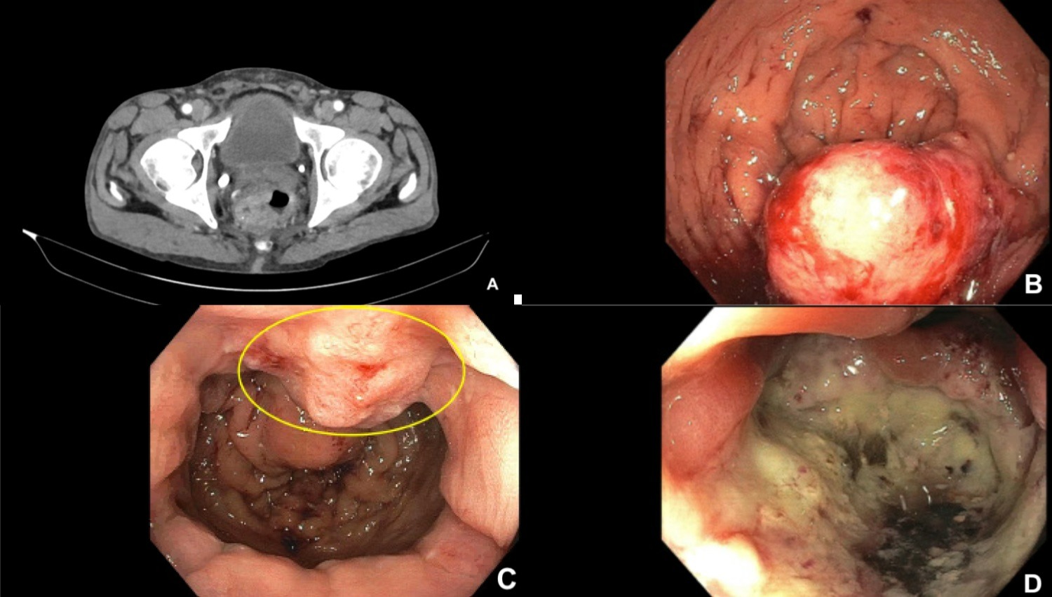
Case Description/Methods: A 59 year-old-male with a history of AIDS presented with bloody diarrhea and fevers. He denied abdominal or rectal pain. Physical exam only revealed mild tenderness of the abdomen. Labs were unremarkable. Gastrointestinal PCR revealed shigella, enteroaggregative E. Coli, enterotoxigenic E. coli, and enteropathogenic E. coli infection. Computed tomography of the abdomen and pelvis revealed pancolitis and left irregular rectal wall thickening, concerning for rectal carcinoma. He was started on IV ciprofloxacin and metronidazole with improvement in symptoms. Colonoscopy revealed a non-obstructing mass in the rectum. Pathology demonstrated high-grade anal intraepithelial lesion (AIN-3) and focally invasive squamous cell carcinoma, with acute and chronically inflamed rectal mucosa. Given the incongruence between colonoscopy and pathology, it was uncertain whether a primary lesion of the rectum with spread to anus existed or if two separate pathologies were present. Repeat colonoscopy to define the anatomic location of the mass revealed a severely ulcerated, friable, rectal lesion, discontinuous from an area of anal inflammation, separated by normal mucosa. Repeat pathology revealed invasive and in situ SCC of the rectum. The patient was referred for chemoradiation with 5-fluorouracil and mitomycin.
Discussion: Primary rectal SCC accounts for 0.01% of all colo-rectal carcinomas. Risk is increased in patients with a history of Human Papilloma virus infection. The diagnosis of primary rectal SCC requires three criteria: (i) exclusion of metastasis, (ii) no squamous‐lined fistulous tract involving the affected rectum, and (iii) exclusion of anal SCC with proximal extension (absence of continuity between the tumor and the normal anal squamous epithelium). Our patient ultimately fulfilled all these criteria. Primary rectal SCC should be considered a differential diagnosis in patients with HIV/AIDS, presenting with bloody diarrhea and a rectal mass.

Disclosures:
Gres Karim, MD1, Frederick Rozenshteyn, MD2, Edward Lung, MD3, Priya Simoes, MBBS4. A0142 - Is It Anal or Rectal? A Rare Case of Squamous Cell Carcinoma, ACG 2022 Annual Scientific Meeting Abstracts. Charlotte, NC: American College of Gastroenterology.
1Mount Sinai Beth Israel, New York, NY; 2Icahn School of Medicine at Mount Sinai Morningside-West, New York, NY; 3Mount Sinai Morningside and Mount Sinai West, New York, NY; 4Mount Sinai West and Morningside Hospital, New York, NY
Introduction: Primary rectal squamous cell carcinomas (SCC) are extremely rare and difficult to distinguish from anal cancers. The majority of rectal SCC are secondary to anal SCC extension and detected at an advanced stage. Although rectal and anal cancers are anatomically close, they are distinct entities with risk factors, different histologic features, patterns of spread, staging systems, and treatment pathways. We present a rare case of a middle-aged man with AIDS who presented with bloody diarrhea, eventually found to have a primary rectal SCC.
Case Description/Methods: A 59 year-old-male with a history of AIDS presented with bloody diarrhea and fevers. He denied abdominal or rectal pain. Physical exam only revealed mild tenderness of the abdomen. Labs were unremarkable. Gastrointestinal PCR revealed shigella, enteroaggregative E. Coli, enterotoxigenic E. coli, and enteropathogenic E. coli infection. Computed tomography of the abdomen and pelvis revealed pancolitis and left irregular rectal wall thickening, concerning for rectal carcinoma. He was started on IV ciprofloxacin and metronidazole with improvement in symptoms. Colonoscopy revealed a non-obstructing mass in the rectum. Pathology demonstrated high-grade anal intraepithelial lesion (AIN-3) and focally invasive squamous cell carcinoma, with acute and chronically inflamed rectal mucosa. Given the incongruence between colonoscopy and pathology, it was uncertain whether a primary lesion of the rectum with spread to anus existed or if two separate pathologies were present. Repeat colonoscopy to define the anatomic location of the mass revealed a severely ulcerated, friable, rectal lesion, discontinuous from an area of anal inflammation, separated by normal mucosa. Repeat pathology revealed invasive and in situ SCC of the rectum. The patient was referred for chemoradiation with 5-fluorouracil and mitomycin.
Discussion: Primary rectal SCC accounts for 0.01% of all colo-rectal carcinomas. Risk is increased in patients with a history of Human Papilloma virus infection. The diagnosis of primary rectal SCC requires three criteria: (i) exclusion of metastasis, (ii) no squamous‐lined fistulous tract involving the affected rectum, and (iii) exclusion of anal SCC with proximal extension (absence of continuity between the tumor and the normal anal squamous epithelium). Our patient ultimately fulfilled all these criteria. Primary rectal SCC should be considered a differential diagnosis in patients with HIV/AIDS, presenting with bloody diarrhea and a rectal mass.

Figure: Figure 1: CT of the abdomen and pelvis (A) revealing an irregular, asymmetric rectal wall thickening centered laterally on the left, highly concerning for rectal carcinoma, in addition to a focal left-sided outpouching from the rectum, suggesting a thick-walled diverticulum. Colonoscopy showing a rectal mass with ulcerated and friable mucosa (B&D). Area of anal inflammation (yellow circle, C) discontinuous from rectal mass with an adjacent small area of normal epithelium.
Disclosures:
Gres Karim indicated no relevant financial relationships.
Frederick Rozenshteyn indicated no relevant financial relationships.
Edward Lung indicated no relevant financial relationships.
Priya Simoes indicated no relevant financial relationships.
Gres Karim, MD1, Frederick Rozenshteyn, MD2, Edward Lung, MD3, Priya Simoes, MBBS4. A0142 - Is It Anal or Rectal? A Rare Case of Squamous Cell Carcinoma, ACG 2022 Annual Scientific Meeting Abstracts. Charlotte, NC: American College of Gastroenterology.
