Back
Poster Session D - Tuesday Morning
D0125 - Rectal Xanthoma: A Rare Clinical Finding
Tuesday, October 25, 2022
10:00 AM – 12:00 PM ET
Location: Crown Ballroom

Joseph Wooten, MD
Temple University Hospital
Philadelphia, PA
Presenting Author(s)
Joseph Wooten, MD1, Randa Hennawy, MD1, Saraswathi Cappelle, DO2, Neena Mohan, MD1
1Temple University Hospital, Philadelphia, PA; 2Lewis Katz School of Medicine at Temple University, Philadelphia, PA
Introduction: Xanthomas are localized lipid deposits within an organ system. They are more commonly found as cutaneous lesions and less likely to appear in the gastrointestinal tract. The incidence of gastrointestinal xanthomas is not well documented, but the vast majority of authors agree they occur more frequently in the stomach. Colonic xanthomas are rare and usually incidental endoscopic findings. There are several case reports that present gastrointestinal xanthomas, however a small subset of these cases are located in the rectum. Here we present a case of rectal xanthoma and discuss its macroscopic and microscopic features.
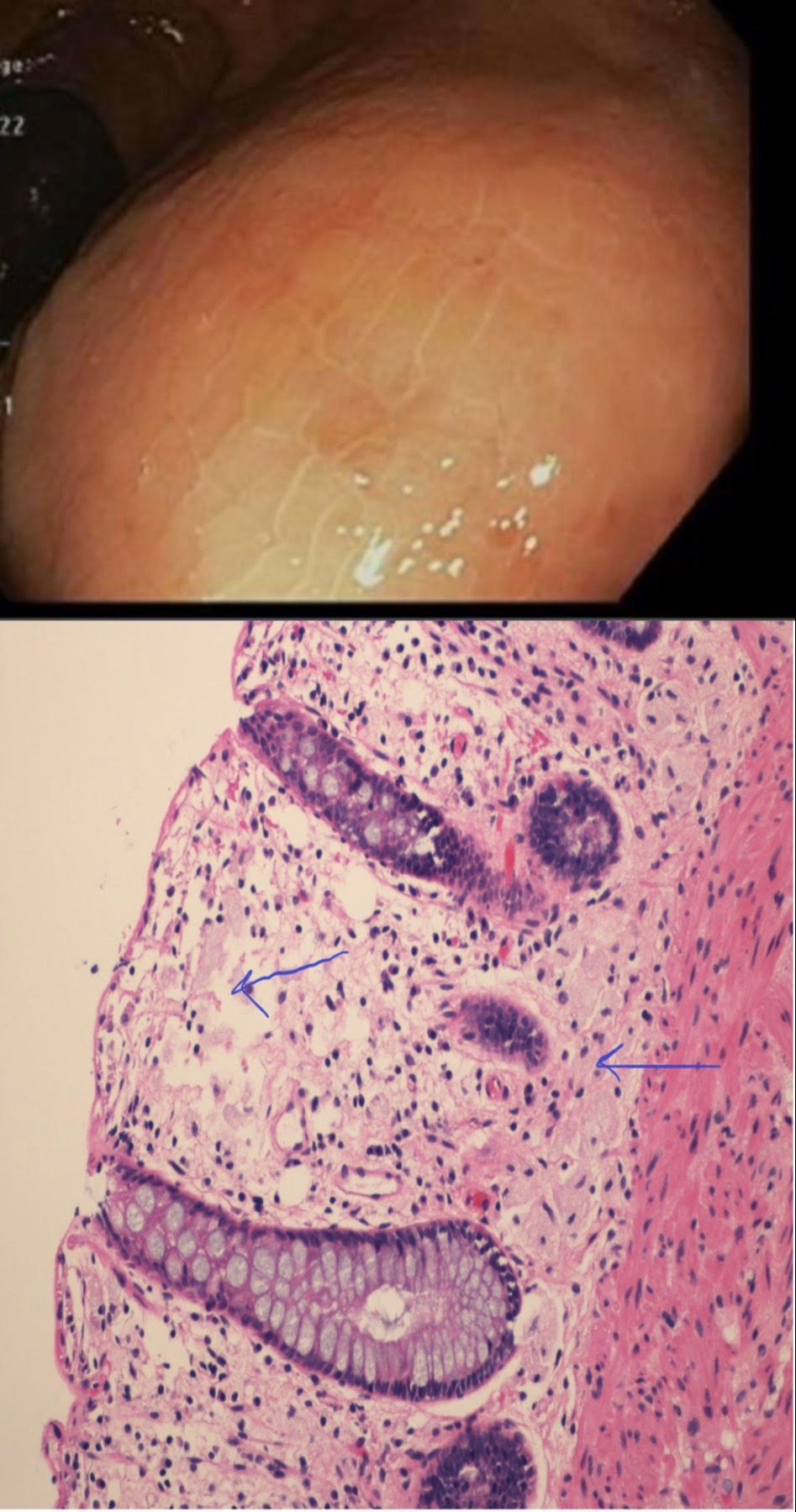
Case Description/Methods: A 55 year old female with a recent diagnosis of atrophic metaplastic autoimmune gastritis presents for colonoscopy. Per chart review, patient had a normal colonoscopy in 2012. During the colonoscopy in 2022, one 10 mm sessile polyp was resected in the cecum; pathology returned as hyperplastic polyp. In the rectum was an area concerning for a submucosal lesion, encompassing about one quarter of the rectal circumference. Overlying it was an area of mucosa with hues of yellow which was biopsied. Pathology showed fragments of colonic mucosa with mild chronic inactive inflammation with histiocytic aggregate and focal surface glandular hyperplasia suggestive of xanthoma. Lipid panel available from 2019 showed LDL 100 with other levels within normal range.
Discussion: Colorectal xanthomas are intestinal lesions with aggregates of lipid laden macrophages called foamy histiocytes. These cells are distributed in the lamina propria, between the colonic glands and muscularis mucosa. Unlike cutaneous xanthomas, gastrointestinal xanthomas are not associated with dyslipidemia. There has been a reported case of colonic xanthomas presenting as submucosal masses in the rectum and sigmoid. In a case series of 28 patients with colorectal xanthomas, 23 were sessile and 5 were pedunculated. Twelve xanthomas appeared reddish in color, 5 were white, and 2 were of a yellow hue. The relationship between colorectal xanthomas and malignancy is unclear. Of the 28 colorectal xanthomas, 4 hyperplastic lesions were found within the xanthoma, while 4 adenomas and 2 adenocarcinomas were found adjacent to xanthomas. Chronic injury is believed to be associated with the cause of colorectal xanthomas. In our case, a sigmoidoscopy with EUS is planned to rule out a submucosal mass. Adding case reports to the literature will help assess potential correlation with polyps and malignancies.

Disclosures:
Joseph Wooten, MD1, Randa Hennawy, MD1, Saraswathi Cappelle, DO2, Neena Mohan, MD1. D0125 - Rectal Xanthoma: A Rare Clinical Finding, ACG 2022 Annual Scientific Meeting Abstracts. Charlotte, NC: American College of Gastroenterology.
1Temple University Hospital, Philadelphia, PA; 2Lewis Katz School of Medicine at Temple University, Philadelphia, PA
Introduction: Xanthomas are localized lipid deposits within an organ system. They are more commonly found as cutaneous lesions and less likely to appear in the gastrointestinal tract. The incidence of gastrointestinal xanthomas is not well documented, but the vast majority of authors agree they occur more frequently in the stomach. Colonic xanthomas are rare and usually incidental endoscopic findings. There are several case reports that present gastrointestinal xanthomas, however a small subset of these cases are located in the rectum. Here we present a case of rectal xanthoma and discuss its macroscopic and microscopic features.
Case Description/Methods: A 55 year old female with a recent diagnosis of atrophic metaplastic autoimmune gastritis presents for colonoscopy. Per chart review, patient had a normal colonoscopy in 2012. During the colonoscopy in 2022, one 10 mm sessile polyp was resected in the cecum; pathology returned as hyperplastic polyp. In the rectum was an area concerning for a submucosal lesion, encompassing about one quarter of the rectal circumference. Overlying it was an area of mucosa with hues of yellow which was biopsied. Pathology showed fragments of colonic mucosa with mild chronic inactive inflammation with histiocytic aggregate and focal surface glandular hyperplasia suggestive of xanthoma. Lipid panel available from 2019 showed LDL 100 with other levels within normal range.
Discussion: Colorectal xanthomas are intestinal lesions with aggregates of lipid laden macrophages called foamy histiocytes. These cells are distributed in the lamina propria, between the colonic glands and muscularis mucosa. Unlike cutaneous xanthomas, gastrointestinal xanthomas are not associated with dyslipidemia. There has been a reported case of colonic xanthomas presenting as submucosal masses in the rectum and sigmoid. In a case series of 28 patients with colorectal xanthomas, 23 were sessile and 5 were pedunculated. Twelve xanthomas appeared reddish in color, 5 were white, and 2 were of a yellow hue. The relationship between colorectal xanthomas and malignancy is unclear. Of the 28 colorectal xanthomas, 4 hyperplastic lesions were found within the xanthoma, while 4 adenomas and 2 adenocarcinomas were found adjacent to xanthomas. Chronic injury is believed to be associated with the cause of colorectal xanthomas. In our case, a sigmoidoscopy with EUS is planned to rule out a submucosal mass. Adding case reports to the literature will help assess potential correlation with polyps and malignancies.

Figure: Figure 1 (top): Rectum with appearance of submucosal lesion and localized area of yellow hue in mucosa
Figure 2 (bottom): Foamy histiocytes confined to the lamina propria.
Figure 2 (bottom): Foamy histiocytes confined to the lamina propria.
Disclosures:
Joseph Wooten indicated no relevant financial relationships.
Randa Hennawy indicated no relevant financial relationships.
Saraswathi Cappelle: Olympus – Consultant.
Neena Mohan indicated no relevant financial relationships.
Neena Mohan — NO DISCLOSURE DATA.
Joseph Wooten, MD1, Randa Hennawy, MD1, Saraswathi Cappelle, DO2, Neena Mohan, MD1. D0125 - Rectal Xanthoma: A Rare Clinical Finding, ACG 2022 Annual Scientific Meeting Abstracts. Charlotte, NC: American College of Gastroenterology.
