Back


Poster Session C - Monday Afternoon
Category: Colon
C0133 - Diffuse Large B Cell Lymphoma Presenting as a Single Colon Polyp With Concurrent Hepatitis B Virus Infection
Monday, October 24, 2022
3:00 PM – 5:00 PM ET
Location: Crown Ballroom

Has Audio

Talia F. Malik, MD
Chicago Medical School at Rosalind Franklin University of Medicine and Science
North Chicago, IL
Presenting Author(s)
Talia F. Malik, MD1, Salma Akram, MD2
1Chicago Medical School at Rosalind Franklin University of Medicine and Science, North Chicago, IL; 2Wright State University Boonshoft School of Medcine, Dayton, OH
Introduction: Primary colonic lymphomas are exceedingly rare accounting for 1% of gastrointestinal lymphomas and 0.2-0.5% of all colorectal malignancies. Diffuse large B-cell lymphoma (DLBCL) is strongly associated with hepatitis B virus (HBV) with a prevalence of 30.2%, however, less is known about its impact on overall disease prognosis. We report a case of colonic DLBCL localized in a polyp in an HBV-infected patient.
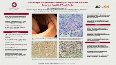
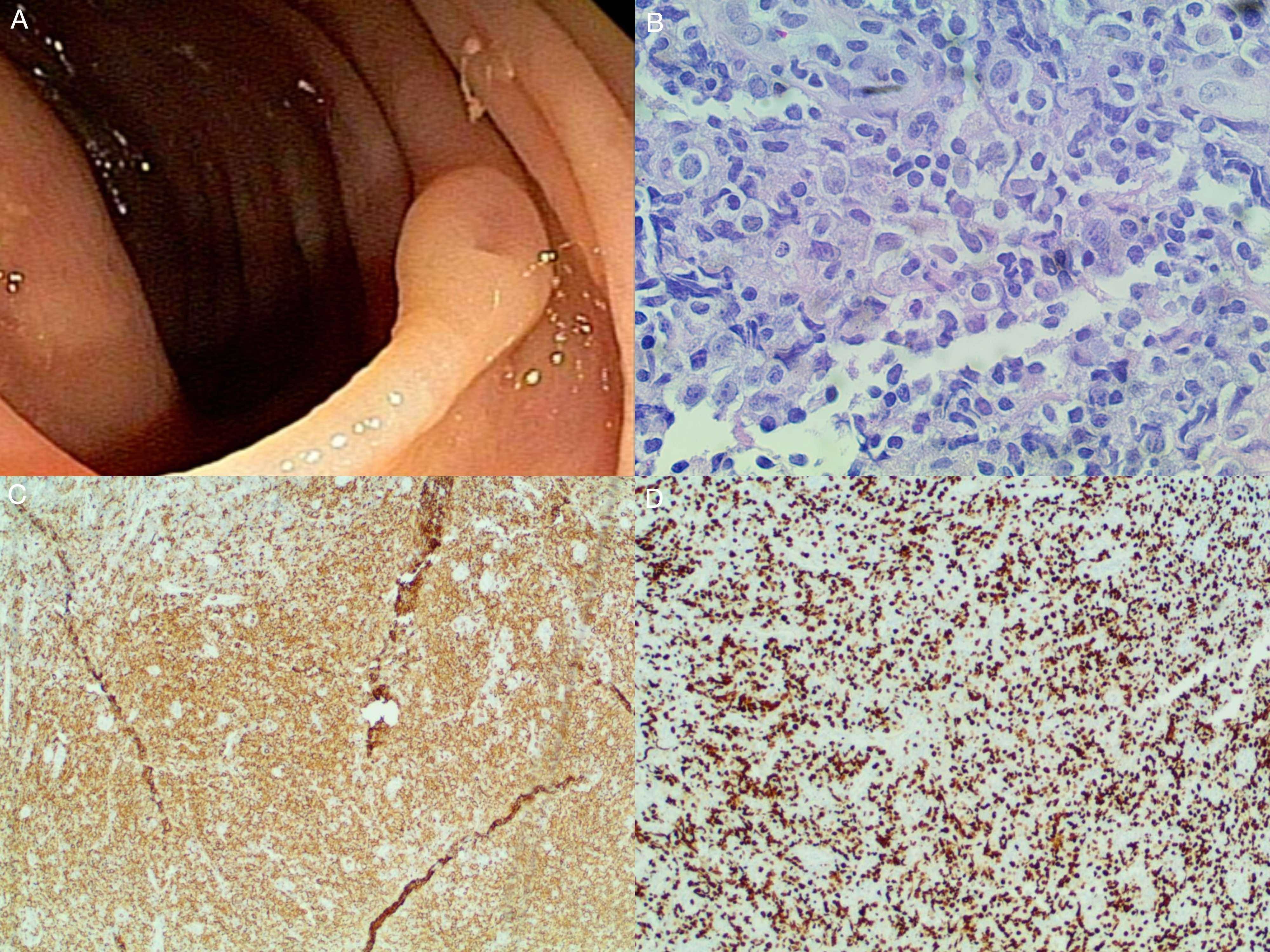
Case Description/Methods: A 50-year-old Asian female with history of untreated HBV infection presented for a screening colonoscopy. She denied any fever, night sweats, fatigue, weight loss, or changes in bowel habits. Physical exam and laboratory workup were largely unremarkable. She was positive for hepatitis B e antigen (HBeAg) and hepatitis B surface antigen (HBsAg), and HBV DNA was 731 IU/ml. Colonoscopy revealed a single sessile 5 mm polyp in the descending colon that was completely resected using a hot polypectomy snare (Figure 1A). Histopathological evaluation revealed the presence of atypical lymphoid cells with prominent nucleoli (Figure 1B). Immunohistochemical analyses revealed lymphocytic cells positive for CD20, PAX5, CD79a, BCL2, MUM1, CD30, and negative for Cyclin D1, BCL6, CD10, ALK, and cMYC (Figure 1C, 1D). She was started on anti-HBV treatment with tenofovir 300 mg daily. A PET scan and bone marrow biopsy and aspirate were negative, consistent with diagnosis of stage I DLBCL. Patient declined chemotherapy. A repeat PET scan and colonoscopy two years later did not show any recurrent disease.
Discussion: Gastrointestinal DLBCL is extremely rare accounting for 1.4% of Non-Hodgkin Lymphomas. It most commonly arises in the stomach, followed by the small intestine and colon. Primary colonic DLBCL usually presents as a fungating mass in the ileocecal region and rarely as a benign-appearing colonic polyp. It is plausible that concurrent HBV infection was associated with the unusual presentation of DLBCL in this asymptomatic patient. This reinforces the importance of a low threshold for endoscopic sampling of any mucosal abnormality during routine screening colonoscopy in HBV-positive patients. This case highlights that complete endoscopic or surgical resection in an early-stage disease could be curative. The impact of concurrent HBV infection on the pathogenesis, disease course, and prognosis of colonic DLBCL remains unclear. It is possible that anti-HBV treatment, especially in HBeAg and HBsAg positive cases, may lead to improved outcomes.

Disclosures:
Talia F. Malik, MD1, Salma Akram, MD2. C0133 - Diffuse Large B Cell Lymphoma Presenting as a Single Colon Polyp With Concurrent Hepatitis B Virus Infection, ACG 2022 Annual Scientific Meeting Abstracts. Charlotte, NC: American College of Gastroenterology.
1Chicago Medical School at Rosalind Franklin University of Medicine and Science, North Chicago, IL; 2Wright State University Boonshoft School of Medcine, Dayton, OH
Introduction: Primary colonic lymphomas are exceedingly rare accounting for 1% of gastrointestinal lymphomas and 0.2-0.5% of all colorectal malignancies. Diffuse large B-cell lymphoma (DLBCL) is strongly associated with hepatitis B virus (HBV) with a prevalence of 30.2%, however, less is known about its impact on overall disease prognosis. We report a case of colonic DLBCL localized in a polyp in an HBV-infected patient.
Case Description/Methods: A 50-year-old Asian female with history of untreated HBV infection presented for a screening colonoscopy. She denied any fever, night sweats, fatigue, weight loss, or changes in bowel habits. Physical exam and laboratory workup were largely unremarkable. She was positive for hepatitis B e antigen (HBeAg) and hepatitis B surface antigen (HBsAg), and HBV DNA was 731 IU/ml. Colonoscopy revealed a single sessile 5 mm polyp in the descending colon that was completely resected using a hot polypectomy snare (Figure 1A). Histopathological evaluation revealed the presence of atypical lymphoid cells with prominent nucleoli (Figure 1B). Immunohistochemical analyses revealed lymphocytic cells positive for CD20, PAX5, CD79a, BCL2, MUM1, CD30, and negative for Cyclin D1, BCL6, CD10, ALK, and cMYC (Figure 1C, 1D). She was started on anti-HBV treatment with tenofovir 300 mg daily. A PET scan and bone marrow biopsy and aspirate were negative, consistent with diagnosis of stage I DLBCL. Patient declined chemotherapy. A repeat PET scan and colonoscopy two years later did not show any recurrent disease.
Discussion: Gastrointestinal DLBCL is extremely rare accounting for 1.4% of Non-Hodgkin Lymphomas. It most commonly arises in the stomach, followed by the small intestine and colon. Primary colonic DLBCL usually presents as a fungating mass in the ileocecal region and rarely as a benign-appearing colonic polyp. It is plausible that concurrent HBV infection was associated with the unusual presentation of DLBCL in this asymptomatic patient. This reinforces the importance of a low threshold for endoscopic sampling of any mucosal abnormality during routine screening colonoscopy in HBV-positive patients. This case highlights that complete endoscopic or surgical resection in an early-stage disease could be curative. The impact of concurrent HBV infection on the pathogenesis, disease course, and prognosis of colonic DLBCL remains unclear. It is possible that anti-HBV treatment, especially in HBeAg and HBsAg positive cases, may lead to improved outcomes.

Figure: Figure 1. (A) Endoscopic image showing a 5 mm sessile polyp in the descending colon. (B) Large atypical lymphoid cells with pleomorphic nuclei, vesicular chromatin, and distinct central nucleoli (H&E, 400x magnification). (C) Atypical lymphoid cells staining positive for CD20 (40x magnification). (D) Atypical lymphoid cells staining positive for MUM1 (40x magnification).
Disclosures:
Talia Malik indicated no relevant financial relationships.
Salma Akram indicated no relevant financial relationships.
Talia F. Malik, MD1, Salma Akram, MD2. C0133 - Diffuse Large B Cell Lymphoma Presenting as a Single Colon Polyp With Concurrent Hepatitis B Virus Infection, ACG 2022 Annual Scientific Meeting Abstracts. Charlotte, NC: American College of Gastroenterology.

