Back


Poster Session C - Monday Afternoon
Category: Colon
C0152 - Disseminated Histoplasmosis Presenting as Right Colon Lesions in HIV-Negative Patients: Report of Two Cases
Monday, October 24, 2022
3:00 PM – 5:00 PM ET
Location: Crown Ballroom

Has Audio

Arturo Suplee Rivera, MD
University of Texas Rio Grande Valley at Doctors Hospital at Renaissance
Edinburg, TX
Presenting Author(s)
Arturo Suplee Rivera, MD, Prateek S. Harne, MBBS, MD, Murthy Badiga, MD, FACG, Asif Zamir, MD, FACG
University of Texas Rio Grande Valley at Doctors Hospital at Renaissance, Edinburg, TX
Introduction: Histoplasma capsulatum is the most common endemic mycosis in the gastrointestinal tract of immunocompromised hosts. However, it can also present in immunocompetent patients. Terminal ileum and cecum are most commonly affected, recognized on endoscopic examination as inflammation, ulcerated lesions, or polypoid masses. In most patients, gastrointestinal manifestation takes a subclinical disease course. We present two cases of immunocompetent patients with colon lesions attributed to disseminated histoplasmosis.
Case Description/Methods:
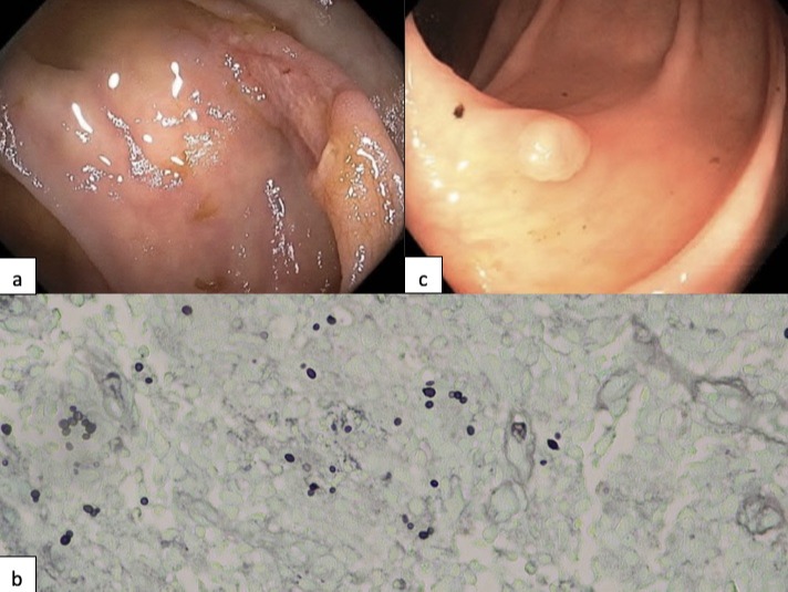
First case is a 62-year-old African American woman admitted to the hospital due to abdominal pain, weight loss and poor oral intake. She was found to be anemic. CT revealed a large retroperitoneal mass with central necrosis suspicious for malignancy. Colonoscopy revealed a cecal ulcer (Image 1a) with pathology reporting mucosal ulceration associated with granulomatous inflammation. GMS and PAS fungal stain showed small ovoid yeast forms, morphologically compatible with histoplasmosis (Image 1b).Biopsy of the retroperitoneal mass was also positive for histoplasma.
Second case is a 54-year-old Hispanic man admitted to the hospital with fluid overload secondary to acute renal failure attributed to post-infectious glomerulonephritis. He was also found to have anemia. Colonoscopy showed a 6 mm polyp in the ascending colon removed by cold snare technique (Image 1c). Pathology reported polyp fragment with macrophages and numerous 2-4 micron non-budding yeast forms best seen on GMS stain, most consistent with histoplasmosis. CT scan showed prominent retroperitoneal lymphadenopathy. Both our patients were started in itraconazole and are being closely followed by the ID service.
Discussion: Disseminated histoplasmosis is usually seen in immunocompromised patients such as those with HIV and medication-induced immunosuppression. GI involvement and symptoms can be diverse. Our first patient presented with a large retroperitoneal mass and cecal ulcer causing her abdominal discomfort. Second patient was much more subtle as histoplasmosis presented as a polypoid lesion. Both patients presented with significant anemia, which prompted endoscopic evaluation. Although the diagnosis of histoplasmosis in immunocompetent patients usually requires a high index of suspicion, gastroenterologists should be familiar with its variable presentations in the GI tract.

Disclosures:
Arturo Suplee Rivera, MD, Prateek S. Harne, MBBS, MD, Murthy Badiga, MD, FACG, Asif Zamir, MD, FACG. C0152 - Disseminated Histoplasmosis Presenting as Right Colon Lesions in HIV-Negative Patients: Report of Two Cases, ACG 2022 Annual Scientific Meeting Abstracts. Charlotte, NC: American College of Gastroenterology.
University of Texas Rio Grande Valley at Doctors Hospital at Renaissance, Edinburg, TX
Introduction: Histoplasma capsulatum is the most common endemic mycosis in the gastrointestinal tract of immunocompromised hosts. However, it can also present in immunocompetent patients. Terminal ileum and cecum are most commonly affected, recognized on endoscopic examination as inflammation, ulcerated lesions, or polypoid masses. In most patients, gastrointestinal manifestation takes a subclinical disease course. We present two cases of immunocompetent patients with colon lesions attributed to disseminated histoplasmosis.
Case Description/Methods:
First case is a 62-year-old African American woman admitted to the hospital due to abdominal pain, weight loss and poor oral intake. She was found to be anemic. CT revealed a large retroperitoneal mass with central necrosis suspicious for malignancy. Colonoscopy revealed a cecal ulcer (Image 1a) with pathology reporting mucosal ulceration associated with granulomatous inflammation. GMS and PAS fungal stain showed small ovoid yeast forms, morphologically compatible with histoplasmosis (Image 1b).Biopsy of the retroperitoneal mass was also positive for histoplasma.
Second case is a 54-year-old Hispanic man admitted to the hospital with fluid overload secondary to acute renal failure attributed to post-infectious glomerulonephritis. He was also found to have anemia. Colonoscopy showed a 6 mm polyp in the ascending colon removed by cold snare technique (Image 1c). Pathology reported polyp fragment with macrophages and numerous 2-4 micron non-budding yeast forms best seen on GMS stain, most consistent with histoplasmosis. CT scan showed prominent retroperitoneal lymphadenopathy. Both our patients were started in itraconazole and are being closely followed by the ID service.
Discussion: Disseminated histoplasmosis is usually seen in immunocompromised patients such as those with HIV and medication-induced immunosuppression. GI involvement and symptoms can be diverse. Our first patient presented with a large retroperitoneal mass and cecal ulcer causing her abdominal discomfort. Second patient was much more subtle as histoplasmosis presented as a polypoid lesion. Both patients presented with significant anemia, which prompted endoscopic evaluation. Although the diagnosis of histoplasmosis in immunocompetent patients usually requires a high index of suspicion, gastroenterologists should be familiar with its variable presentations in the GI tract.

Figure: Image 1: a) Histoplasma presenting as cecal ulcer. b) GMS stain showing Histoplasma. c) Polypoid lesion positive for Histoplasma.
Disclosures:
Arturo Suplee Rivera indicated no relevant financial relationships.
Prateek Harne indicated no relevant financial relationships.
Murthy Badiga indicated no relevant financial relationships.
Asif Zamir indicated no relevant financial relationships.
Arturo Suplee Rivera, MD, Prateek S. Harne, MBBS, MD, Murthy Badiga, MD, FACG, Asif Zamir, MD, FACG. C0152 - Disseminated Histoplasmosis Presenting as Right Colon Lesions in HIV-Negative Patients: Report of Two Cases, ACG 2022 Annual Scientific Meeting Abstracts. Charlotte, NC: American College of Gastroenterology.
