Back


Poster Session C - Monday Afternoon
Category: Esophagus
C0220 - Co-Localization of Immunoglobulin G4 (IgG4) and Milk Proteins Is Associated With Eosinophilic Esophagitis Disease Activity
Monday, October 24, 2022
3:00 PM – 5:00 PM ET
Location: Crown Ballroom

Has Audio

Jonathan Medernach, DO
UVA Health
Charlottesville, VA
Presenting Author(s)
Award: Presidential Poster Award
Jonathan Medernach, DO1, Rung-chi Li, DO, PhD1, Elaine Etter, PhD1, Shyam Raghavan, MD1, Emily Noonan, BS1, Larry Borish, MD1, Barrett Barnes, MD1, Thomas Platts-Mills, MD, PhD, FRS1, Bryan G. Sauer, MD, MSc2, Emily McGowan, MD, PhD1
1UVA Health, Charlottesville, VA; 2University of Virginia, Charlottesville, VA
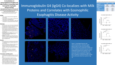
Introduction: Eosinophilic esophagitis is a chronic disease of the esophagus predominantly triggered by food antigens but diagnostic tests to identify food triggers in individual patients do not yet exist. Recent studies have shown that immunoglobulin G4 (IgG4) co-localizes with food proteins in the esophageal tissue of eosinophilic esophagitis (EoE) patients. Knowing that cow’s milk is the main food trigger of EoE, we hypothesized that co-localization with IgG4 is associated with EoE disease activity.
Methods: We performed a case control study nested within the prospective University of Virginia (UVA) EoE Cohort. Fifteen patients were selected from the UVA EoE Cohort (10 EoE patients and 5 controls). Esophageal biopsies were obtained at the time of each endoscopy. We examined paired samples from individuals with a) active EoE and remission on swallowed steroids, b) active EoE and remission on diet elimination, c) non-EoE controls (n=5 in each group). Immunofluorescence was performed using primary antibodies directed against IgG4 and milk proteins (Bos d 4,5,8). Images were captured using a Leica confocal microscope. Co-localization was determined using Pearson’s Correlation Coefficient on Imaris imaging software. Between-group comparisons were made using Mann-Whitney U test on GraphPad Prism software.
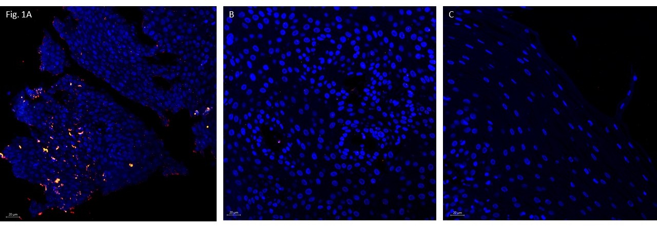
Results: Immunofluorescence staining was performed on 75 esophageal biopsies (15 patients, ages 19-41 yo, 53% male). There was significantly more IgG4-milk protein co-localization in patients with active EoE compared to remission samples in the same patients (p=0.02, p=0.002, and p=0.002 for Bos d 4, 5, and 8, respectively). There was no significant difference in co-localization between remission samples and controls (p=0.57, p=0.21, p=0.74 for Bos d 4, 5, and 8, respectively). Median co-localization levels in active EoE patients were also significantly higher than non-EoE controls. Notably, all five patients in remission on swallowed steroids still demonstrated co-localization but at a lesser intensity compared to active samples and was not statistically significant (p=0.07).
Discussion: Our findings demonstrate that IgG4 co-localization with milk proteins is associated with disease activity in EoE. Whether these deposits contribute to the underlying inflammation of EoE, and whether IgG4 co-localization could be used to identify specific food triggers in EoE patients remains unknown and warrants further study.

Disclosures:
Jonathan Medernach, DO1, Rung-chi Li, DO, PhD1, Elaine Etter, PhD1, Shyam Raghavan, MD1, Emily Noonan, BS1, Larry Borish, MD1, Barrett Barnes, MD1, Thomas Platts-Mills, MD, PhD, FRS1, Bryan G. Sauer, MD, MSc2, Emily McGowan, MD, PhD1. C0220 - Co-Localization of Immunoglobulin G4 (IgG4) and Milk Proteins Is Associated With Eosinophilic Esophagitis Disease Activity, ACG 2022 Annual Scientific Meeting Abstracts. Charlotte, NC: American College of Gastroenterology.
Jonathan Medernach, DO1, Rung-chi Li, DO, PhD1, Elaine Etter, PhD1, Shyam Raghavan, MD1, Emily Noonan, BS1, Larry Borish, MD1, Barrett Barnes, MD1, Thomas Platts-Mills, MD, PhD, FRS1, Bryan G. Sauer, MD, MSc2, Emily McGowan, MD, PhD1
1UVA Health, Charlottesville, VA; 2University of Virginia, Charlottesville, VA
Introduction: Eosinophilic esophagitis is a chronic disease of the esophagus predominantly triggered by food antigens but diagnostic tests to identify food triggers in individual patients do not yet exist. Recent studies have shown that immunoglobulin G4 (IgG4) co-localizes with food proteins in the esophageal tissue of eosinophilic esophagitis (EoE) patients. Knowing that cow’s milk is the main food trigger of EoE, we hypothesized that co-localization with IgG4 is associated with EoE disease activity.
Methods: We performed a case control study nested within the prospective University of Virginia (UVA) EoE Cohort. Fifteen patients were selected from the UVA EoE Cohort (10 EoE patients and 5 controls). Esophageal biopsies were obtained at the time of each endoscopy. We examined paired samples from individuals with a) active EoE and remission on swallowed steroids, b) active EoE and remission on diet elimination, c) non-EoE controls (n=5 in each group). Immunofluorescence was performed using primary antibodies directed against IgG4 and milk proteins (Bos d 4,5,8). Images were captured using a Leica confocal microscope. Co-localization was determined using Pearson’s Correlation Coefficient on Imaris imaging software. Between-group comparisons were made using Mann-Whitney U test on GraphPad Prism software.
Results: Immunofluorescence staining was performed on 75 esophageal biopsies (15 patients, ages 19-41 yo, 53% male). There was significantly more IgG4-milk protein co-localization in patients with active EoE compared to remission samples in the same patients (p=0.02, p=0.002, and p=0.002 for Bos d 4, 5, and 8, respectively). There was no significant difference in co-localization between remission samples and controls (p=0.57, p=0.21, p=0.74 for Bos d 4, 5, and 8, respectively). Median co-localization levels in active EoE patients were also significantly higher than non-EoE controls. Notably, all five patients in remission on swallowed steroids still demonstrated co-localization but at a lesser intensity compared to active samples and was not statistically significant (p=0.07).
Discussion: Our findings demonstrate that IgG4 co-localization with milk proteins is associated with disease activity in EoE. Whether these deposits contribute to the underlying inflammation of EoE, and whether IgG4 co-localization could be used to identify specific food triggers in EoE patients remains unknown and warrants further study.

Figure: Figure 1: Esophageal biopsies with immunofluorescence staining for IgG4 (green), Bos d 5 (red), and nuclei (blue). (A) Active EoE and co-localization of IgG4 and Bos d 5 (yellow). (B) Paired remission sample (swallowed steroids) from patient in fig. 1A. (C) Control patient consuming dairy
Disclosures:
Jonathan Medernach indicated no relevant financial relationships.
Rung-chi Li indicated no relevant financial relationships.
Elaine Etter indicated no relevant financial relationships.
Shyam Raghavan indicated no relevant financial relationships.
Emily Noonan indicated no relevant financial relationships.
Larry Borish indicated no relevant financial relationships.
Barrett Barnes indicated no relevant financial relationships.
Thomas Platts-Mills: NIH (R37-AI20565) – Grant/Research Support.
Bryan Sauer: Advarra – Advisor or Review Panel Member. Takeda – Consultant.
Emily McGowan: Regeneron Pharmaceuticals – Grant/Research Support.
Jonathan Medernach, DO1, Rung-chi Li, DO, PhD1, Elaine Etter, PhD1, Shyam Raghavan, MD1, Emily Noonan, BS1, Larry Borish, MD1, Barrett Barnes, MD1, Thomas Platts-Mills, MD, PhD, FRS1, Bryan G. Sauer, MD, MSc2, Emily McGowan, MD, PhD1. C0220 - Co-Localization of Immunoglobulin G4 (IgG4) and Milk Proteins Is Associated With Eosinophilic Esophagitis Disease Activity, ACG 2022 Annual Scientific Meeting Abstracts. Charlotte, NC: American College of Gastroenterology.

