Back


Poster Session C - Monday Afternoon
Category: Esophagus
C0251 - A Rare Cause of Esophageal Varices
Monday, October 24, 2022
3:00 PM – 5:00 PM ET
Location: Crown Ballroom

Has Audio

Jason J. John, MD
University of South Florida Health
Tampa, FL
Presenting Author(s)
Jason J. John, MD1, Koushik R. Reddy, MD2, Wojciech Blonski, MD, PhD2
1University of South Florida Health, Tampa, FL; 2James A. Haley VA Hospital, Tampa, FL
Introduction: Esophageal varices (EV) are classified as downhill or uphill varices. Uphill varices are common and found in the distal esophagus that develop secondary to portal hypertension. Downhill esophageal varices (DEV) are rare and found in the proximal and mid esophagus and most commonly develop from obstructed venous blood flow in the superior vena cava (SVC).
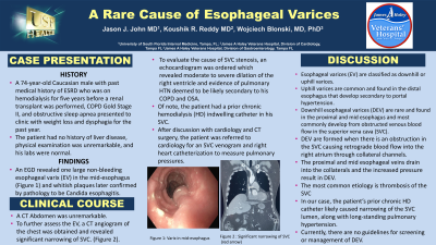
Case Description/Methods: A 74-year-old Caucasian male with past medical history of ESRD who was on hemodialysis for five years before a renal transplant was performed, COPD Gold Stage II, and obstructive sleep apnea presented to clinic with weight loss and dysphagia for the past year. The patient had no history of liver disease, physical examination was unremarkable, and his labs were normal. An esophagogastroduodenoscopy (EGD) revealed one large non-bleeding esophageal varix (EV) in the mid-esophagus and whitish plaques later confirmed by pathology to be Candida esophagitis. The patient was placed on IV micafungin and his dysphagia resolved. A CT Abdomen showed a normal liver and spleen. To further assess the DEV, a CT angiogram of the chest was obtained and revealed significant narrowing of SVC. (Figure 1). To evaluate the cause of SVC stenosis, an echocardiogram was ordered which revealed moderate to severe dilation of the right ventricle and evidence of pulmonary hypertension deemed to be likely secondary to his COPD and OSA. Of note, the patient had a prior chronic hemodialysis (HD) indwelling catheter in his SVC. After discussion with cardiology and CT surgery, the patient was referred to cardiology for an SVC venogram and right heart catheterization to measure pulmonary pressures.
Discussion: DEV are formed when there is an obstruction in the SVC causing retrograde blood flow into the right atrium through collateral channels. The proximal and mid esophageal veins drain into the collaterals and the increased pressure result in DEV. The most common etiology is thrombosis of the SVC, but DEV can also be caused by severe pulmonary hypertension, thyroid tumors, and complications with HD catheters. In our case, the patient’s prior chronic HD catheter likely caused narrowing of the SVC lumen, along with long-standing pulmonary hypertension. Currently, there are no guidelines for screening or management of DEV. Physicians should be aware of unusual causes of varices and the management of DEV involves a multidisciplinary approach to treat the underlying cause of the SVC obstruction.

Disclosures:
Jason J. John, MD1, Koushik R. Reddy, MD2, Wojciech Blonski, MD, PhD2. C0251 - A Rare Cause of Esophageal Varices, ACG 2022 Annual Scientific Meeting Abstracts. Charlotte, NC: American College of Gastroenterology.
1University of South Florida Health, Tampa, FL; 2James A. Haley VA Hospital, Tampa, FL
Introduction: Esophageal varices (EV) are classified as downhill or uphill varices. Uphill varices are common and found in the distal esophagus that develop secondary to portal hypertension. Downhill esophageal varices (DEV) are rare and found in the proximal and mid esophagus and most commonly develop from obstructed venous blood flow in the superior vena cava (SVC).
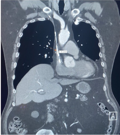
Case Description/Methods: A 74-year-old Caucasian male with past medical history of ESRD who was on hemodialysis for five years before a renal transplant was performed, COPD Gold Stage II, and obstructive sleep apnea presented to clinic with weight loss and dysphagia for the past year. The patient had no history of liver disease, physical examination was unremarkable, and his labs were normal. An esophagogastroduodenoscopy (EGD) revealed one large non-bleeding esophageal varix (EV) in the mid-esophagus and whitish plaques later confirmed by pathology to be Candida esophagitis. The patient was placed on IV micafungin and his dysphagia resolved. A CT Abdomen showed a normal liver and spleen. To further assess the DEV, a CT angiogram of the chest was obtained and revealed significant narrowing of SVC. (Figure 1). To evaluate the cause of SVC stenosis, an echocardiogram was ordered which revealed moderate to severe dilation of the right ventricle and evidence of pulmonary hypertension deemed to be likely secondary to his COPD and OSA. Of note, the patient had a prior chronic hemodialysis (HD) indwelling catheter in his SVC. After discussion with cardiology and CT surgery, the patient was referred to cardiology for an SVC venogram and right heart catheterization to measure pulmonary pressures.
Discussion: DEV are formed when there is an obstruction in the SVC causing retrograde blood flow into the right atrium through collateral channels. The proximal and mid esophageal veins drain into the collaterals and the increased pressure result in DEV. The most common etiology is thrombosis of the SVC, but DEV can also be caused by severe pulmonary hypertension, thyroid tumors, and complications with HD catheters. In our case, the patient’s prior chronic HD catheter likely caused narrowing of the SVC lumen, along with long-standing pulmonary hypertension. Currently, there are no guidelines for screening or management of DEV. Physicians should be aware of unusual causes of varices and the management of DEV involves a multidisciplinary approach to treat the underlying cause of the SVC obstruction.

Figure: Figure 1: Significant narrowing of SVC (red arrow) on CT chest
Disclosures:
Jason John indicated no relevant financial relationships.
Koushik Reddy indicated no relevant financial relationships.
Wojciech Blonski indicated no relevant financial relationships.
Jason J. John, MD1, Koushik R. Reddy, MD2, Wojciech Blonski, MD, PhD2. C0251 - A Rare Cause of Esophageal Varices, ACG 2022 Annual Scientific Meeting Abstracts. Charlotte, NC: American College of Gastroenterology.
