Back
Poster Session A - Sunday Afternoon
Category: Biliary/Pancreas
A0058 - A Case of Adenomyomatosis of the Gallbladder: Evaluating Malignant Potential
Sunday, October 23, 2022
5:00 PM – 7:00 PM ET
Location: Crown Ballroom
- VP
Viraaj S. Pannu, MBBS
Jersey Shore University Medical Center
Apex, North Carolina
Presenting Author(s)
Viraaj S. Pannu, MBBS1, Mihir Odak, MD2, Steven Douedi, MD3, Kameron Tavakolian, MD3, Justin A. Ilagan, DO4, Swapnil Patel, MD3
1Jersey Shore University Medical Center, Apex, NC; 2Jersey Shore University Medical Center, Monroe Township, NJ; 3Jersey Shore University Medical Center, Neptune, NJ; 4Jersey Shore University Medical Center, Tinton Falls, NJ
Introduction: Adenomyomatosis of the gallbladder(GA) is a rare pathology involving muscularis growth in the gallbladder(GB). It is associated with an increased risk of gallbladder malignancy. While usually incidentally found post-resection or on abdominal imaging. We present a case of a 42-year-old female who was found to have GA on right upper quadrant ultrasound, in the hopes of encouraging prompt prophylactic removal in such cases due to a risk of progression to malignancy.
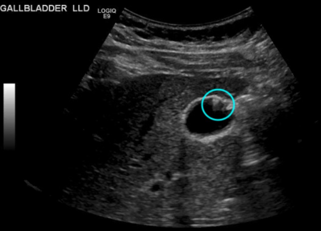
Case Description/Methods: A 42-year-old female presented with right upper quadrant pain of 1-year duration. The pain was intermittent, associated with nausea and some degree of constipation, which resolved with fiber supplements. She had no change in the nature of her stool nor any bloating or excessive belching. She also reported no correlation of her pain with food intake, although did report a decreased appetite during this time. Laboratory data was unremarkable. Due to persistent, yet intermittent pain, a right upper quadrant ultrasound was obtained, which revealed focal adenomyomatosis involving the anterior gallbladder wall. The patient was subsequently referred for laparoscopic cholecystectomy with the general surgery team and on follow-up, has been symptom-free since her procedure.
Discussion: GA is a benign incidental finding in as many as 9% of cholecystectomy samples. GA is thought to develop due to persistently elevated intrabiliary pressures, ultimately resulting in a hyperplastic muscularis layer and polyps [4]. Though there is no consensus on the progression of GA to adenocarcinoma of the GB, GA is associated with a less than 5% five-year mortality if left untreated[3]. A study by Ootani et al also described a 6% incidence of GB adenocarcinoma in patients with segmental type GA as opposed to only 3% in patients without GA[3]. Given the association of malignancies with chronic inflammation, GA may render an environment favorable for cancerous growth. With this case study we hope to advocate for the need for more research on the possible association of GB adenocarcinoma in patients with GA, particularly the segmental type and if there are any benefits from prophylactic cholecystectomy in patients with GA found incidentally.

Disclosures:
Viraaj S. Pannu, MBBS1, Mihir Odak, MD2, Steven Douedi, MD3, Kameron Tavakolian, MD3, Justin A. Ilagan, DO4, Swapnil Patel, MD3. A0058 - A Case of Adenomyomatosis of the Gallbladder: Evaluating Malignant Potential, ACG 2022 Annual Scientific Meeting Abstracts. Charlotte, NC: American College of Gastroenterology.
1Jersey Shore University Medical Center, Apex, NC; 2Jersey Shore University Medical Center, Monroe Township, NJ; 3Jersey Shore University Medical Center, Neptune, NJ; 4Jersey Shore University Medical Center, Tinton Falls, NJ
Introduction: Adenomyomatosis of the gallbladder(GA) is a rare pathology involving muscularis growth in the gallbladder(GB). It is associated with an increased risk of gallbladder malignancy. While usually incidentally found post-resection or on abdominal imaging. We present a case of a 42-year-old female who was found to have GA on right upper quadrant ultrasound, in the hopes of encouraging prompt prophylactic removal in such cases due to a risk of progression to malignancy.
Case Description/Methods: A 42-year-old female presented with right upper quadrant pain of 1-year duration. The pain was intermittent, associated with nausea and some degree of constipation, which resolved with fiber supplements. She had no change in the nature of her stool nor any bloating or excessive belching. She also reported no correlation of her pain with food intake, although did report a decreased appetite during this time. Laboratory data was unremarkable. Due to persistent, yet intermittent pain, a right upper quadrant ultrasound was obtained, which revealed focal adenomyomatosis involving the anterior gallbladder wall. The patient was subsequently referred for laparoscopic cholecystectomy with the general surgery team and on follow-up, has been symptom-free since her procedure.
Discussion: GA is a benign incidental finding in as many as 9% of cholecystectomy samples. GA is thought to develop due to persistently elevated intrabiliary pressures, ultimately resulting in a hyperplastic muscularis layer and polyps [4]. Though there is no consensus on the progression of GA to adenocarcinoma of the GB, GA is associated with a less than 5% five-year mortality if left untreated[3]. A study by Ootani et al also described a 6% incidence of GB adenocarcinoma in patients with segmental type GA as opposed to only 3% in patients without GA[3]. Given the association of malignancies with chronic inflammation, GA may render an environment favorable for cancerous growth. With this case study we hope to advocate for the need for more research on the possible association of GB adenocarcinoma in patients with GA, particularly the segmental type and if there are any benefits from prophylactic cholecystectomy in patients with GA found incidentally.

Figure: Right Upper Quadrant Ultrasound showing likely adenomyomatosis of the Gallbladder wall
Disclosures:
Viraaj Pannu indicated no relevant financial relationships.
Mihir Odak indicated no relevant financial relationships.
Steven Douedi indicated no relevant financial relationships.
Kameron Tavakolian indicated no relevant financial relationships.
Justin Ilagan indicated no relevant financial relationships.
Swapnil Patel indicated no relevant financial relationships.
Viraaj S. Pannu, MBBS1, Mihir Odak, MD2, Steven Douedi, MD3, Kameron Tavakolian, MD3, Justin A. Ilagan, DO4, Swapnil Patel, MD3. A0058 - A Case of Adenomyomatosis of the Gallbladder: Evaluating Malignant Potential, ACG 2022 Annual Scientific Meeting Abstracts. Charlotte, NC: American College of Gastroenterology.
