Back


Poster Session C - Monday Afternoon
Category: Interventional Endoscopy
C0467 - Hepatic Tuberculoma Caused by Mycobacterium Fortuitum: A Rare Case Diagnosed by EUS-FNA
Monday, October 24, 2022
3:00 PM – 5:00 PM ET
Location: Crown Ballroom

Has Audio

Yue-Sai Jao, MD
University of Puerto Rico Medical Sciences Campus
San Juan, Puerto Rico
Presenting Author(s)
Yue-Sai Jao, MD1, Victor J. Carlo Chevere, MD2, Lemuel Martinez, MD3, Carlos G. Micames, MD4
1University of Puerto Rico Medical Sciences Campus, San Juan, Puerto Rico; 2Puerto Rico Pathology Cytology, Hato Rey, Puerto Rico; 3Doctors' Center Hospital, Inc., Manati, Puerto Rico; 4Hospital Bella Vista, Mayaguez, Puerto Rico
Introduction: Mycobacterium fortuitum is a type of nontuberculous mycobacteria belonging to a group classified as Rapidly Growing Mycobacteria (RGM), along with Mycobacterium abscessus and Mycobacterium chelonae. M. fortuitum is predominantly associated with skin, soft tissue, and surgical-wound infections. Catheter-related sepsis and disseminated disease can also occur. Isolated liver involvement by atypical mycobacterial infection is exceedingly rare. Here we describe a case of M. fortuitum presenting as an asymptomatic liver mass.
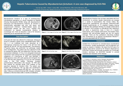
Case Description/Methods: A 67-year-old male was referred for endoscopic ultrasound (EUS) to evaluate a gastric body subepithelial lesion (SEL) and a 3 cm incidental liver mass. He was asymptomatic. Percutaneous CT-guided biopsy was performed, but pathologic results were nondiagnostic. Patient was thus referred for EUS to evaluate the gastric SEL and determine if the liver mass represented metastasis. A 14 by 4 mm elongated intramural hypoechoic mass arising from the muscularis propia highly suggestive of a leiomyoma or GIST was identified. A heterogeneous mass with cystic spaces and echogenic foci in the left liver lobe measuring 27 by 20 mm was sampled using a 22g FNA needle. Pathology revealed granulomatous inflammation with necrosis. An AFB stain was positive for acid fast bacilli. PCR analysis of the specimen detected M. fortuitum DNA. Patient was treated with levofloxacin 750 mg daily plus trimethoprim and sulfamethoxazole (Septra DS) 800 mg - 160 mg twice daily. Follow-up contrast-enhanced MRI revealed complete resolution of liver lesion after 3 months of antibiotics.
Discussion: Isolated liver involvement by nontuberculous mycobacteria is uncommon, usually asymptomatic, and its diagnosis can easily be missed or mistaken. We report the first case of a hepatic tuberculoma caused by M. fortuitum diagnosed by EUS-FNA. Precise and accurate recognition by a minimally invasive technique via EUS-FNA resulted in prompt treatment and resolution of the disease.
Disclosures:
Yue-Sai Jao, MD1, Victor J. Carlo Chevere, MD2, Lemuel Martinez, MD3, Carlos G. Micames, MD4. C0467 - Hepatic Tuberculoma Caused by Mycobacterium Fortuitum: A Rare Case Diagnosed by EUS-FNA, ACG 2022 Annual Scientific Meeting Abstracts. Charlotte, NC: American College of Gastroenterology.
1University of Puerto Rico Medical Sciences Campus, San Juan, Puerto Rico; 2Puerto Rico Pathology Cytology, Hato Rey, Puerto Rico; 3Doctors' Center Hospital, Inc., Manati, Puerto Rico; 4Hospital Bella Vista, Mayaguez, Puerto Rico
Introduction: Mycobacterium fortuitum is a type of nontuberculous mycobacteria belonging to a group classified as Rapidly Growing Mycobacteria (RGM), along with Mycobacterium abscessus and Mycobacterium chelonae. M. fortuitum is predominantly associated with skin, soft tissue, and surgical-wound infections. Catheter-related sepsis and disseminated disease can also occur. Isolated liver involvement by atypical mycobacterial infection is exceedingly rare. Here we describe a case of M. fortuitum presenting as an asymptomatic liver mass.
Case Description/Methods: A 67-year-old male was referred for endoscopic ultrasound (EUS) to evaluate a gastric body subepithelial lesion (SEL) and a 3 cm incidental liver mass. He was asymptomatic. Percutaneous CT-guided biopsy was performed, but pathologic results were nondiagnostic. Patient was thus referred for EUS to evaluate the gastric SEL and determine if the liver mass represented metastasis. A 14 by 4 mm elongated intramural hypoechoic mass arising from the muscularis propia highly suggestive of a leiomyoma or GIST was identified. A heterogeneous mass with cystic spaces and echogenic foci in the left liver lobe measuring 27 by 20 mm was sampled using a 22g FNA needle. Pathology revealed granulomatous inflammation with necrosis. An AFB stain was positive for acid fast bacilli. PCR analysis of the specimen detected M. fortuitum DNA. Patient was treated with levofloxacin 750 mg daily plus trimethoprim and sulfamethoxazole (Septra DS) 800 mg - 160 mg twice daily. Follow-up contrast-enhanced MRI revealed complete resolution of liver lesion after 3 months of antibiotics.
Discussion: Isolated liver involvement by nontuberculous mycobacteria is uncommon, usually asymptomatic, and its diagnosis can easily be missed or mistaken. We report the first case of a hepatic tuberculoma caused by M. fortuitum diagnosed by EUS-FNA. Precise and accurate recognition by a minimally invasive technique via EUS-FNA resulted in prompt treatment and resolution of the disease.
Disclosures:
Yue-Sai Jao indicated no relevant financial relationships.
Victor Carlo Chevere indicated no relevant financial relationships.
Lemuel Martinez: Cubist Pharmaceuticals – Speakers Bureau.
Carlos Micames: Boston Scientific – Consultant. Bristol Myers Squibb – Advisor or Review Panel Member. Janssen – Advisor or Review Panel Member.
Yue-Sai Jao, MD1, Victor J. Carlo Chevere, MD2, Lemuel Martinez, MD3, Carlos G. Micames, MD4. C0467 - Hepatic Tuberculoma Caused by Mycobacterium Fortuitum: A Rare Case Diagnosed by EUS-FNA, ACG 2022 Annual Scientific Meeting Abstracts. Charlotte, NC: American College of Gastroenterology.
