Back


Poster Session B - Monday Morning
Category: Esophagus
B0229 - A Rare Case of Metastatic Muscle Invasive Bladder Cancer to the Esophagus
Monday, October 24, 2022
10:00 AM – 12:00 PM ET
Location: Crown Ballroom

Has Audio

Rafael Rivera Sepulveda, MD
University of South Florida Morsani College of Medicine
Tampa, FL
Presenting Author(s)
Award: Presidential Poster Award
Rafael Rivera Sepulveda, MD1, Charles P. Cavalaris, MD1, Ali Zakaria, MD2, Rene D. Gomez-Esquivel, MD3
1University of South Florida Morsani College of Medicine, Tampa, FL; 2University of South Florida, Tampa, FL; 3USF Health Morsani College of Medicine, Tampa, FL
Introduction: Muscle invasive bladder cancer (MIBC) spreading to the esophagus is very rare. Here we report a case of a severe esophageal stricture caused by metastatic spread of MIBC.
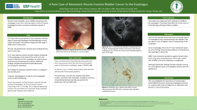
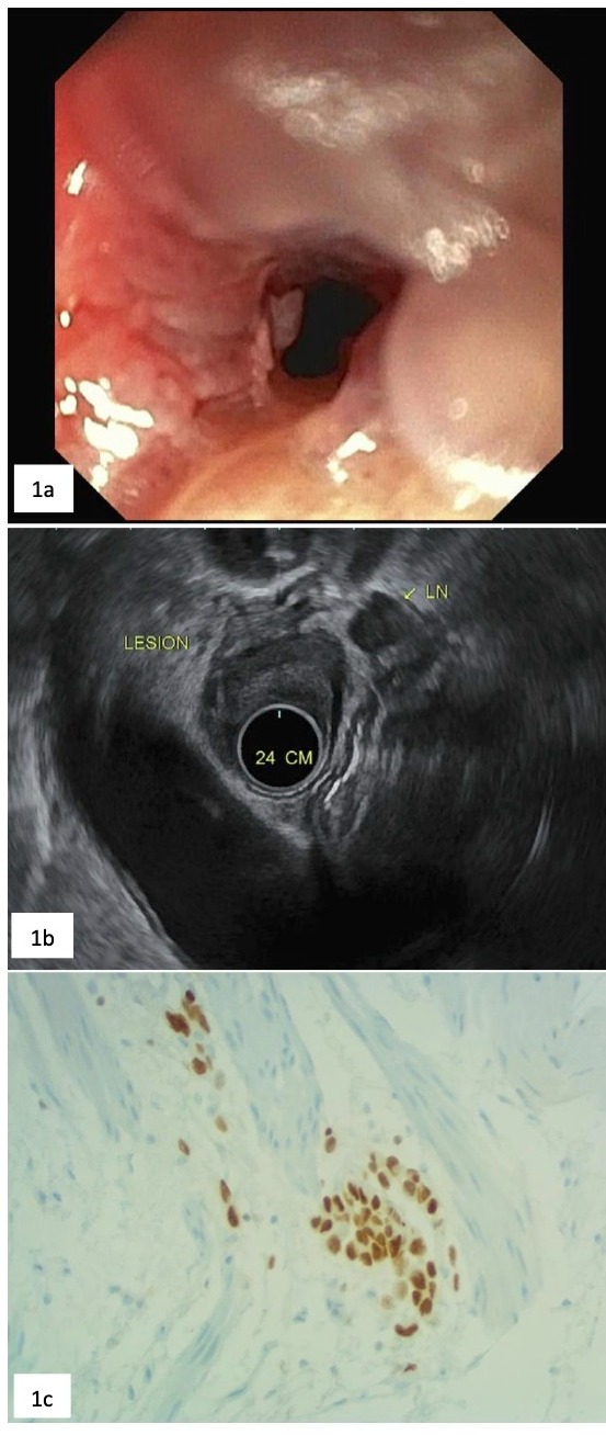
Case Description/Methods: A 57-year-old man presented to our institution with four days of worsening dysphagia and odynophagia to both solids and liquids. He had been diagnosed with stage IV MIBC three months prior. Of note, the patient had a 20-pack-year smoking history, quit 15 years ago. A CT chest without contrast showed nodular thickening of the esophagus at the level of the aortic arch and superior distension of the esophagus as evidenced by a small amount of layering oral contrast. Additional findings included mediastinal lymphadenopathy concerning for metastasis. No findings of metastasis had been seen at a staging CT two months prior. A barium esophagogram revealed a mid-esophageal stricture with shouldering. Direct visualization via EGD showed a severely stenotic section of esophagus, at 25 cm from the incisors, measuring 5 mm x 1 cm (1a). This malignant appearing stricture was only able to be traversed using a pediatric gastroscope. Biopsies were obtained. Then a limited radial echoendoscopy was performed from the proximal end of the stricture and showed a mass in the thoracic esophagus, stage uT3N2Mx (1b). An 18 mm x 12.3 cm fully covered metal stent was then placed under fluoroscopic guidance. Histopathology revealed rare, atypical cells within muscle, consistent with metastatic urothelial carcinoma. Immunohistochemical staining was positive for pankeratin, CK7 and GATA (1c). The patient was diagnosed with metastasis of MIBC to the esophagus. The patient died after the fourth cycle of chemotherapy with gemcitabine-cisplatin.
Discussion: Over the last decade, there have been increasing reports of esophageal cancer metastasizing to the bladder. Here we report a case of bladder cancer metastasizing to the esophagus. To our knowledge, this is the 2nd ever mentioned report of such a finding. Documented incidence of metastasis to the esophagus is about 6%. MIBC most commonly spreads to regional lymph nodes, bone, lung, liver, and peritoneum. The 5-year survival rate of MIBC is 6% once metastasis is established. Although endoscopic findings showed irregular mucosa, not all malignant causes of esophageal stenosis have this appearance. This case highlights the importance of maintaining a broad differential when assessing esophageal strictures on a patient with known malignancy as metastasis may present in uncommon places.

Disclosures:
Rafael Rivera Sepulveda, MD1, Charles P. Cavalaris, MD1, Ali Zakaria, MD2, Rene D. Gomez-Esquivel, MD3. B0229 - A Rare Case of Metastatic Muscle Invasive Bladder Cancer to the Esophagus, ACG 2022 Annual Scientific Meeting Abstracts. Charlotte, NC: American College of Gastroenterology.
Rafael Rivera Sepulveda, MD1, Charles P. Cavalaris, MD1, Ali Zakaria, MD2, Rene D. Gomez-Esquivel, MD3
1University of South Florida Morsani College of Medicine, Tampa, FL; 2University of South Florida, Tampa, FL; 3USF Health Morsani College of Medicine, Tampa, FL
Introduction: Muscle invasive bladder cancer (MIBC) spreading to the esophagus is very rare. Here we report a case of a severe esophageal stricture caused by metastatic spread of MIBC.
Case Description/Methods: A 57-year-old man presented to our institution with four days of worsening dysphagia and odynophagia to both solids and liquids. He had been diagnosed with stage IV MIBC three months prior. Of note, the patient had a 20-pack-year smoking history, quit 15 years ago. A CT chest without contrast showed nodular thickening of the esophagus at the level of the aortic arch and superior distension of the esophagus as evidenced by a small amount of layering oral contrast. Additional findings included mediastinal lymphadenopathy concerning for metastasis. No findings of metastasis had been seen at a staging CT two months prior. A barium esophagogram revealed a mid-esophageal stricture with shouldering. Direct visualization via EGD showed a severely stenotic section of esophagus, at 25 cm from the incisors, measuring 5 mm x 1 cm (1a). This malignant appearing stricture was only able to be traversed using a pediatric gastroscope. Biopsies were obtained. Then a limited radial echoendoscopy was performed from the proximal end of the stricture and showed a mass in the thoracic esophagus, stage uT3N2Mx (1b). An 18 mm x 12.3 cm fully covered metal stent was then placed under fluoroscopic guidance. Histopathology revealed rare, atypical cells within muscle, consistent with metastatic urothelial carcinoma. Immunohistochemical staining was positive for pankeratin, CK7 and GATA (1c). The patient was diagnosed with metastasis of MIBC to the esophagus. The patient died after the fourth cycle of chemotherapy with gemcitabine-cisplatin.
Discussion: Over the last decade, there have been increasing reports of esophageal cancer metastasizing to the bladder. Here we report a case of bladder cancer metastasizing to the esophagus. To our knowledge, this is the 2nd ever mentioned report of such a finding. Documented incidence of metastasis to the esophagus is about 6%. MIBC most commonly spreads to regional lymph nodes, bone, lung, liver, and peritoneum. The 5-year survival rate of MIBC is 6% once metastasis is established. Although endoscopic findings showed irregular mucosa, not all malignant causes of esophageal stenosis have this appearance. This case highlights the importance of maintaining a broad differential when assessing esophageal strictures on a patient with known malignancy as metastasis may present in uncommon places.

Figure: 1a. Malignant appearing intrinsic stenosis seen on EGD measuring 5mm (inner diameter) x 1 cm (in length).
1b. EUS: A mass that was partially circumferential with irregular endosonographic borders was found in the thoracic esophagus at 24 cm from the incisors and lymphadenopathy is seen on the right.
1c. Pathology: Rare, atypical cells within muscle, immunoreactive for GATA stain, consistent with metastatic urothelial carcinoma.
Disclosures:
Rafael Rivera Sepulveda indicated no relevant financial relationships.
Charles Cavalaris indicated no relevant financial relationships.
Ali Zakaria indicated no relevant financial relationships.
Rene Gomez-Esquivel indicated no relevant financial relationships.
Rafael Rivera Sepulveda, MD1, Charles P. Cavalaris, MD1, Ali Zakaria, MD2, Rene D. Gomez-Esquivel, MD3. B0229 - A Rare Case of Metastatic Muscle Invasive Bladder Cancer to the Esophagus, ACG 2022 Annual Scientific Meeting Abstracts. Charlotte, NC: American College of Gastroenterology.

