Back


Poster Session A - Sunday Afternoon
Category: Colon
A0162 - Looks Aren't Everything: A Rare Case of Metastatic Colonic Neuroendocrine Tumor
Sunday, October 23, 2022
5:00 PM – 7:00 PM ET
Location: Crown Ballroom

Has Audio

Harsh Patel, MD, MPH
NewYork Presbyterian Brooklyn Methodist Hospital
Brooklyn, NY
Presenting Author(s)
Harsh Patel, MD, MPH1, Dean Rizzi, MD2, Samuel Quintero, MD3, Ofem Ajah, MD1
1NewYork Presbyterian Brooklyn Methodist Hospital, Brooklyn, NY; 2New York Presbyterian Brooklyn Methodist, Brooklyn, NY; 3NYP Brooklyn Methodist Hospital, Brooklyn, NY
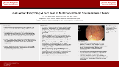
Introduction: Gastrointestinal neuroendocrine tumors (NETs) are rare, slow growing tumors with distinct histopathological characteristics. Endoscopically, they appear as sessile, flat polypoid lesions, making them difficult to distinguish from pathologically benign polyps. They can be found anywhere within the gastrointestinal tract with the terminal ileum being the most common location. Severity of disease varies based on size, macroscopic ulcerations, and hyperemia of mucosal surfaces. NETs of the colon and rectum are extremely rare and account for only 1% of all colorectal malignancies. Patients typically remain asymptomatic until the tumor is large enough to cause obstruction or abdominal pain and is diagnosed during routine colonoscopy.
Case Description/Methods: We present a 63-year-old male with a past medical history of hypertension, hyperlipidemia, and parathyroidectomy, who on colonoscopy in 2019 had three benign-appearing polyps showing mixed hyperplastic and tubular adenoma morphology. Three years later, repeat colonoscopy revealed five sessile polyps of varying sizes from 2 mm to 6 mm, where all were completely resected. Pathology showed 4 tubular adenomas and a 2-millimeter low grade (WHO 2010 Grade 1) well-differentiated neuroendocrine tumor located in the sigmoid colon. The latter exhibited typical findings of NETs with a trabecular growth pattern, round nuclei, and a “salt and pepper” chromatin. Ki-67 proliferative index was 1%. Tumor cells were positive for synaptophysin, and chromogranin, and negative for CK7, CK20 and CDX2. PET scan demonstrated intensely DOTATATE avid mural thickening at the duodenal bulb and proximal second portion of the duodenum, along with uptake in liver, and pulmonary nodules, with osseous lesions suspicious for metastatic disease. Repeat colonoscopy two months later was unrevealing without evidence of neuroendocrine tumor.
Discussion: Colorectal NETs are rapidly progressive with a poor prognosis compared to colorectal adenocarcinoma. Our case demonstrates that low grade, well differentiated NETs of the colon can undergo rapid high-grade transformation within a short interval between colonoscopies. When considering NETs without known metastatic disease, lesions that are amenable to endoscopic resection may be treated with endoscopic intervention and surveillance alone. Given rapid progression of disease and predilection for metastasis, our case highlights the need for early detection of NETs with interval screening colonoscopies to decrease morbidity and mortality.

Disclosures:
Harsh Patel, MD, MPH1, Dean Rizzi, MD2, Samuel Quintero, MD3, Ofem Ajah, MD1. A0162 - Looks Aren't Everything: A Rare Case of Metastatic Colonic Neuroendocrine Tumor, ACG 2022 Annual Scientific Meeting Abstracts. Charlotte, NC: American College of Gastroenterology.
1NewYork Presbyterian Brooklyn Methodist Hospital, Brooklyn, NY; 2New York Presbyterian Brooklyn Methodist, Brooklyn, NY; 3NYP Brooklyn Methodist Hospital, Brooklyn, NY
Introduction: Gastrointestinal neuroendocrine tumors (NETs) are rare, slow growing tumors with distinct histopathological characteristics. Endoscopically, they appear as sessile, flat polypoid lesions, making them difficult to distinguish from pathologically benign polyps. They can be found anywhere within the gastrointestinal tract with the terminal ileum being the most common location. Severity of disease varies based on size, macroscopic ulcerations, and hyperemia of mucosal surfaces. NETs of the colon and rectum are extremely rare and account for only 1% of all colorectal malignancies. Patients typically remain asymptomatic until the tumor is large enough to cause obstruction or abdominal pain and is diagnosed during routine colonoscopy.
Case Description/Methods: We present a 63-year-old male with a past medical history of hypertension, hyperlipidemia, and parathyroidectomy, who on colonoscopy in 2019 had three benign-appearing polyps showing mixed hyperplastic and tubular adenoma morphology. Three years later, repeat colonoscopy revealed five sessile polyps of varying sizes from 2 mm to 6 mm, where all were completely resected. Pathology showed 4 tubular adenomas and a 2-millimeter low grade (WHO 2010 Grade 1) well-differentiated neuroendocrine tumor located in the sigmoid colon. The latter exhibited typical findings of NETs with a trabecular growth pattern, round nuclei, and a “salt and pepper” chromatin. Ki-67 proliferative index was 1%. Tumor cells were positive for synaptophysin, and chromogranin, and negative for CK7, CK20 and CDX2. PET scan demonstrated intensely DOTATATE avid mural thickening at the duodenal bulb and proximal second portion of the duodenum, along with uptake in liver, and pulmonary nodules, with osseous lesions suspicious for metastatic disease. Repeat colonoscopy two months later was unrevealing without evidence of neuroendocrine tumor.
Discussion: Colorectal NETs are rapidly progressive with a poor prognosis compared to colorectal adenocarcinoma. Our case demonstrates that low grade, well differentiated NETs of the colon can undergo rapid high-grade transformation within a short interval between colonoscopies. When considering NETs without known metastatic disease, lesions that are amenable to endoscopic resection may be treated with endoscopic intervention and surveillance alone. Given rapid progression of disease and predilection for metastasis, our case highlights the need for early detection of NETs with interval screening colonoscopies to decrease morbidity and mortality.

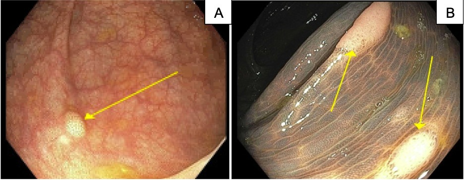
Figure: Image A: 3 mm polyp located in the sigmoid colon. Appears sessile and flat with no malignant appearing lesion. Image B: two polyps located in the transverse colon, 3 mm and 5 mm in size, which endoscopically appear similar to image A. Pathology is consistent with tubular adenoma.
Disclosures:
Harsh Patel indicated no relevant financial relationships.
Dean Rizzi indicated no relevant financial relationships.
Samuel Quintero indicated no relevant financial relationships.
Ofem Ajah indicated no relevant financial relationships.
Harsh Patel, MD, MPH1, Dean Rizzi, MD2, Samuel Quintero, MD3, Ofem Ajah, MD1. A0162 - Looks Aren't Everything: A Rare Case of Metastatic Colonic Neuroendocrine Tumor, ACG 2022 Annual Scientific Meeting Abstracts. Charlotte, NC: American College of Gastroenterology.
