Back


Poster Session A - Sunday Afternoon
Category: Functional Bowel Disease
A0261 - Sclerosing Mesenteritis: A Case Report in a Young Female
Sunday, October 23, 2022
5:00 PM – 7:00 PM ET
Location: Crown Ballroom

Has Audio

Kyle L. Marrache, DO
Abington-Jefferson Health
Abington, Pennsylvania
Presenting Author(s)
Kyle L. Marrache, DO1, Stephanie Tzarnas, MD2
1Abington-Jefferson Health, Abington, PA; 2Abington-Jefferson Hospital, Abington, PA
Introduction: Sclerosing Mesenteritis (SM) is a rare fibrotic inflammatory condition of mesenteric adipose tissue. It is of clinical significance since it appears to carry a five times higher risk of malignancy. In this case report we present a female with both clinical symptoms and radiographic findings suggestive of SM.
Case Description/Methods: A 58-year-old female presented to the resident clinic with the complaint of epigastric pain. On exam she had abdominal distention and tenderness over the epigastrium. Shortly after evaluation in the clinic the patient presented to the ED twice in two weeks with worsening abdominal pain where she got a CT scan both times demonstrating generalized haziness and interval enlargement of mesenteric lymphadenopathy.
Colonoscopy and EGD for her ongoing abdominal pain which were both unremarkable. Duodenal and stomach biopsies were normal. AST, ALT, CRP, ESR, and CBC were all unremarkable. MRI of the pelvis as well as MR enterography was performed and this showed unchanged haziness of the mesentery but found the lymph nodes were not enlarged. The patient was seen by oncology who agreed that her lymphadenopathy had largely resolved, and it was unlikely she had an underlying malignancy.
Surgery was consulted to evaluate for biopsy but felt given the clinical picture of SM was clear it was not warranted.
The patient was seen in the clinic again and started on a regiment of colchicine. She currently has follow-up scheduled.
Discussion: Risk factors for SM are abnormal post-surgical healing, autoimmune mediated, and malignancy. Our patient did have prior abdominal surgery but no other risk factor. Studies have shown a connection with a variety of malignancies, but no statistically significant correlation. The interval enlargement in lymph nodes noted on our patient was concerning but this had resolved on MRI.
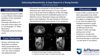
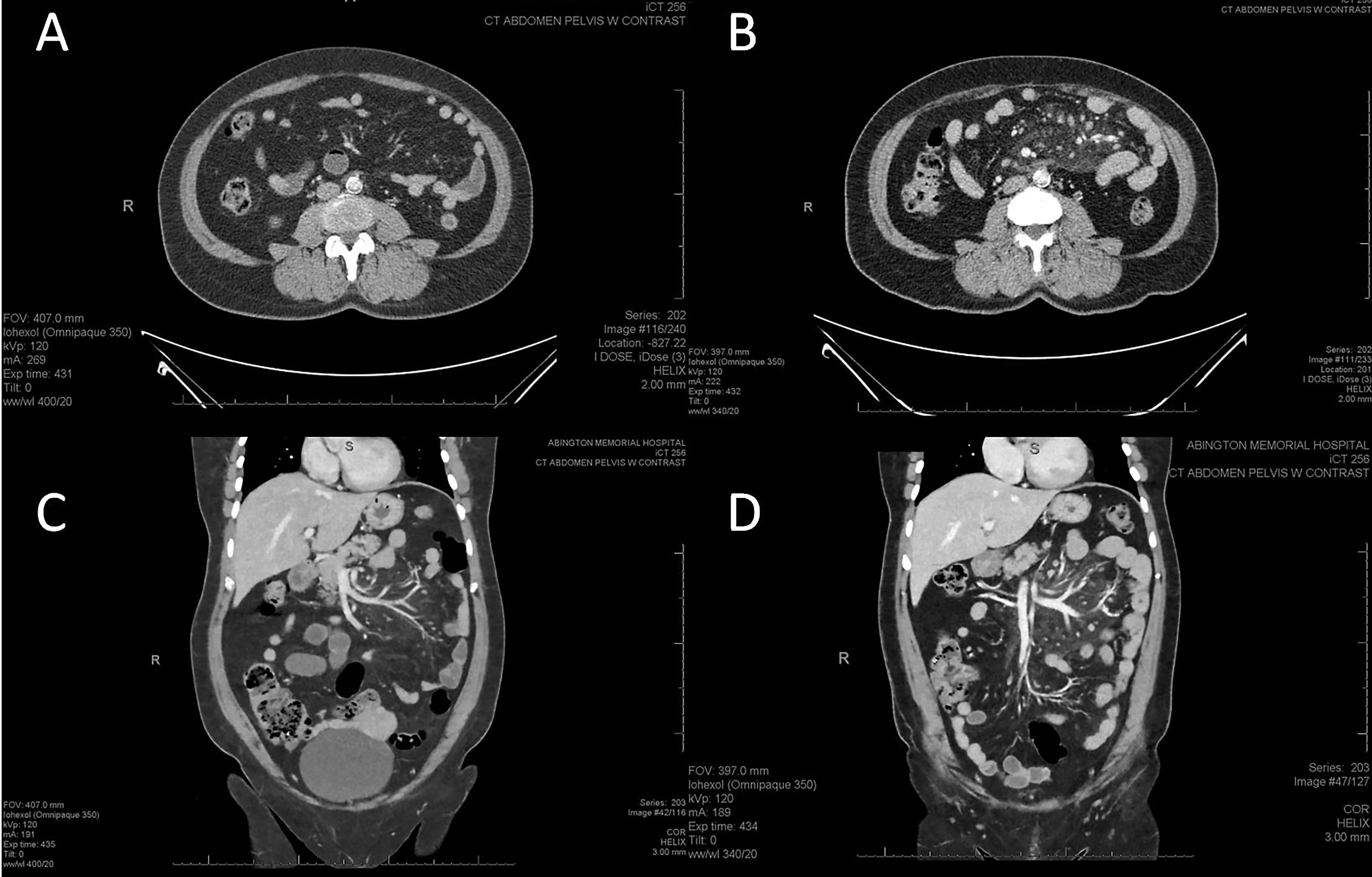
SM is frequently found incidentally on CT scan. Findings suggestive of SM on CT are a “fat ring sign” or “pseudo-capsule” formation. More rarely “misty mesentery” can be seen which is the generalized haziness seen in our patient (fig 1).
Confirmation of SM is by biopsy. In our patient it was felt that the imaging was consistent enough to not warrant a biopsy. Treatment includes prednisone or colchicine. We will continue to follow our patient and see if she has a response to the colchicine with the possibility of starting prednisone in the future if there is no response.

Disclosures:
Kyle L. Marrache, DO1, Stephanie Tzarnas, MD2. A0261 - Sclerosing Mesenteritis: A Case Report in a Young Female, ACG 2022 Annual Scientific Meeting Abstracts. Charlotte, NC: American College of Gastroenterology.
1Abington-Jefferson Health, Abington, PA; 2Abington-Jefferson Hospital, Abington, PA
Introduction: Sclerosing Mesenteritis (SM) is a rare fibrotic inflammatory condition of mesenteric adipose tissue. It is of clinical significance since it appears to carry a five times higher risk of malignancy. In this case report we present a female with both clinical symptoms and radiographic findings suggestive of SM.
Case Description/Methods: A 58-year-old female presented to the resident clinic with the complaint of epigastric pain. On exam she had abdominal distention and tenderness over the epigastrium. Shortly after evaluation in the clinic the patient presented to the ED twice in two weeks with worsening abdominal pain where she got a CT scan both times demonstrating generalized haziness and interval enlargement of mesenteric lymphadenopathy.
Colonoscopy and EGD for her ongoing abdominal pain which were both unremarkable. Duodenal and stomach biopsies were normal. AST, ALT, CRP, ESR, and CBC were all unremarkable. MRI of the pelvis as well as MR enterography was performed and this showed unchanged haziness of the mesentery but found the lymph nodes were not enlarged. The patient was seen by oncology who agreed that her lymphadenopathy had largely resolved, and it was unlikely she had an underlying malignancy.
Surgery was consulted to evaluate for biopsy but felt given the clinical picture of SM was clear it was not warranted.
The patient was seen in the clinic again and started on a regiment of colchicine. She currently has follow-up scheduled.
Discussion: Risk factors for SM are abnormal post-surgical healing, autoimmune mediated, and malignancy. Our patient did have prior abdominal surgery but no other risk factor. Studies have shown a connection with a variety of malignancies, but no statistically significant correlation. The interval enlargement in lymph nodes noted on our patient was concerning but this had resolved on MRI.
SM is frequently found incidentally on CT scan. Findings suggestive of SM on CT are a “fat ring sign” or “pseudo-capsule” formation. More rarely “misty mesentery” can be seen which is the generalized haziness seen in our patient (fig 1).
Confirmation of SM is by biopsy. In our patient it was felt that the imaging was consistent enough to not warrant a biopsy. Treatment includes prednisone or colchicine. We will continue to follow our patient and see if she has a response to the colchicine with the possibility of starting prednisone in the future if there is no response.

Figure: Fig. 1: A: Axial cut from Aug. B: Axial cut from Sep. C: Coronal cut from Aug. D: Coronal cut from Sep
Disclosures:
Kyle Marrache indicated no relevant financial relationships.
Stephanie Tzarnas indicated no relevant financial relationships.
Kyle L. Marrache, DO1, Stephanie Tzarnas, MD2. A0261 - Sclerosing Mesenteritis: A Case Report in a Young Female, ACG 2022 Annual Scientific Meeting Abstracts. Charlotte, NC: American College of Gastroenterology.
