Back


Poster Session C - Monday Afternoon
Category: Stomach
C0717 - Extensive Gastric Mucormycosis in a Patient With Newly Diagnosed Diffuse Large B-Cell Lymphoma of the Stomach
Monday, October 24, 2022
3:00 PM – 5:00 PM ET
Location: Crown Ballroom

Has Audio

Jae Whan Keum, MD
Montefiore Wakefield Hospital
Bronx, New York
Presenting Author(s)
Jae Whan Keum, MD1, Christopher Andrade, MD1, Hilary Hertan, MD, FACG2
1Montefiore Wakefield Hospital, Bronx, NY; 2Montefiore Medical Center, Bronx, NY
Introduction: Non-Hodgkin’s lymphoma (NHL) affects the gastrointestinal tract in 10 to 15 percent of the cases. Mucormycosis in the gastrointestinal tract is rare and may cause severe infection in immunocompromised patients with hematological malignancy. We present a case of newly diagnosed diffuse large B-cell lymphoma (DLBCL) with malignant masses in the stomach and extensive ulcerations with mucormycosis.
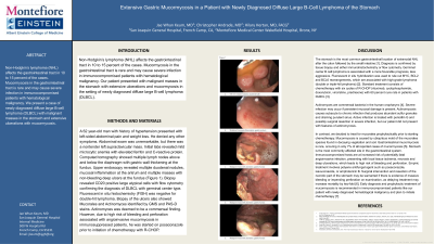
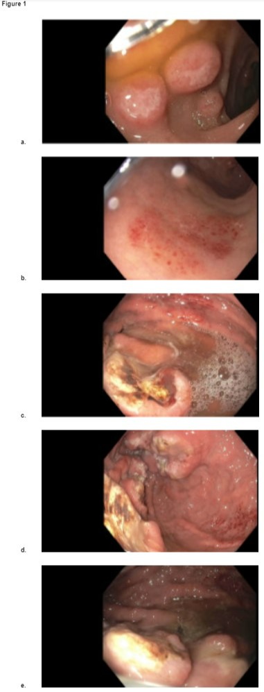
Case Description/Methods: A 62 year-old man with a history of hypertension presented with left abdominal pain and weight loss, associated with decreased appetite. Abdominal exam unremarkable, but there was a nontender left supraclavicular mass. Initial labs revealed mild microcytic anemia with elevated ferritin and C-reactive protein. Computed tomography showed multiple lymphadenopathy above and below the diaphragm with gastric wall thickening at the fundus. Upper endoscopy revealed multiple duodenal nodules, mucosal inflammation at the antrum and multiple masses with non-bleeding deep ulcers at the fundus (Figure 1). Biopsy revealed CD20 positive large atypical cells with flow cytometry confirming the diagnosis of DLBCL with germinal center type. Fluorescent in situ histochemistry (FISH) was negative for double-hit lymphoma. Biopsy of the ulcers also showed Mucorales identified by GMS and PAS-D stains. Patient was started on posaconazole prior to initiation of chemotherapy with R-CHOP.
Discussion: The stomach is the most common gastrointestinal location of extranodal NHL. Diagnosis is confirmed by immunohistochemistry or flow cytometry. Germinal center B cell (GCB) is associated with a favorable prognosis. FISH is used to rule out double or triple-hit high-grade lymphoma with MYC and BCL2 and/or BCL6 rearrangements. Standard treatment consists of six cycles of R-CHOP (rituximab, cyclophosphamide, doxorubicin, vincristine, prednisone) with 60 percent cure rate in patients with DLBCL.
Mucormycosis is caused by mold found in decaying vegetation and soil. Immunocompromised hosts are at risk of potentially fatal angioinvasive infection with local ischemia, necrosis and deep ulcerations. Gastrointestinal mucormycosis occurs in only 7% of reported cases, most commonly affecting the stomach. Empiric treatment with polyene antifungal is necessary. Surgical resection may be needed if there is massive bleeding or impending perforation. Delaying treatment increases mortality by two-fold. Early diagnosis and treatment of mucormycosis is imperative for successful outcome.

Disclosures:
Jae Whan Keum, MD1, Christopher Andrade, MD1, Hilary Hertan, MD, FACG2. C0717 - Extensive Gastric Mucormycosis in a Patient With Newly Diagnosed Diffuse Large B-Cell Lymphoma of the Stomach, ACG 2022 Annual Scientific Meeting Abstracts. Charlotte, NC: American College of Gastroenterology.
1Montefiore Wakefield Hospital, Bronx, NY; 2Montefiore Medical Center, Bronx, NY
Introduction: Non-Hodgkin’s lymphoma (NHL) affects the gastrointestinal tract in 10 to 15 percent of the cases. Mucormycosis in the gastrointestinal tract is rare and may cause severe infection in immunocompromised patients with hematological malignancy. We present a case of newly diagnosed diffuse large B-cell lymphoma (DLBCL) with malignant masses in the stomach and extensive ulcerations with mucormycosis.
Case Description/Methods: A 62 year-old man with a history of hypertension presented with left abdominal pain and weight loss, associated with decreased appetite. Abdominal exam unremarkable, but there was a nontender left supraclavicular mass. Initial labs revealed mild microcytic anemia with elevated ferritin and C-reactive protein. Computed tomography showed multiple lymphadenopathy above and below the diaphragm with gastric wall thickening at the fundus. Upper endoscopy revealed multiple duodenal nodules, mucosal inflammation at the antrum and multiple masses with non-bleeding deep ulcers at the fundus (Figure 1). Biopsy revealed CD20 positive large atypical cells with flow cytometry confirming the diagnosis of DLBCL with germinal center type. Fluorescent in situ histochemistry (FISH) was negative for double-hit lymphoma. Biopsy of the ulcers also showed Mucorales identified by GMS and PAS-D stains. Patient was started on posaconazole prior to initiation of chemotherapy with R-CHOP.
Discussion: The stomach is the most common gastrointestinal location of extranodal NHL. Diagnosis is confirmed by immunohistochemistry or flow cytometry. Germinal center B cell (GCB) is associated with a favorable prognosis. FISH is used to rule out double or triple-hit high-grade lymphoma with MYC and BCL2 and/or BCL6 rearrangements. Standard treatment consists of six cycles of R-CHOP (rituximab, cyclophosphamide, doxorubicin, vincristine, prednisone) with 60 percent cure rate in patients with DLBCL.
Mucormycosis is caused by mold found in decaying vegetation and soil. Immunocompromised hosts are at risk of potentially fatal angioinvasive infection with local ischemia, necrosis and deep ulcerations. Gastrointestinal mucormycosis occurs in only 7% of reported cases, most commonly affecting the stomach. Empiric treatment with polyene antifungal is necessary. Surgical resection may be needed if there is massive bleeding or impending perforation. Delaying treatment increases mortality by two-fold. Early diagnosis and treatment of mucormycosis is imperative for successful outcome.

Figure: Figure 1. Endoscopic findings in the stomach; a. 5-8 mm nodular masses at the duodenal bulb; b. Scattered mucosal inflammation at the gastric antrum; c, d & e. Malignant mass with ulceration at the gastric fundus.
Disclosures:
Jae Whan Keum indicated no relevant financial relationships.
Christopher Andrade indicated no relevant financial relationships.
Hilary Hertan indicated no relevant financial relationships.
Jae Whan Keum, MD1, Christopher Andrade, MD1, Hilary Hertan, MD, FACG2. C0717 - Extensive Gastric Mucormycosis in a Patient With Newly Diagnosed Diffuse Large B-Cell Lymphoma of the Stomach, ACG 2022 Annual Scientific Meeting Abstracts. Charlotte, NC: American College of Gastroenterology.
