Back


Poster Session A - Sunday Afternoon
Category: Colon
A0160 - Cecal Bascule: A Rare Cause of Large Bowel Obstruction
Sunday, October 23, 2022
5:00 PM – 7:00 PM ET
Location: Crown Ballroom

Has Audio

Jonathan E. Selzman, DO
University of Texas Health Science Center
San Antonio, TX
Presenting Author(s)
Jonathan E. Selzman, DO1, Mahesh Gajendran, MD, MPH2, Eric Smith, MD3, Chandraprakash Umapathy, MD4
1University of Texas Health Science Center , San Antonio, TX; 2UTHSCSA, San Antonio, TX; 3BSW- Round Rock, Round Rock, TX; 4University of Texas Health San Antonio, San Antonio, TX
Introduction: Cecal bascule is a rare form of cecal volvulus, which is characterized by an anterior and superiorly displaced cecum in turn causing large bowel obstruction. It accounts for 0.01% of adult large bowel obstructions. Here we present a case of cecal bascule in a cirrhotic patient.
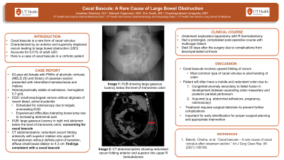
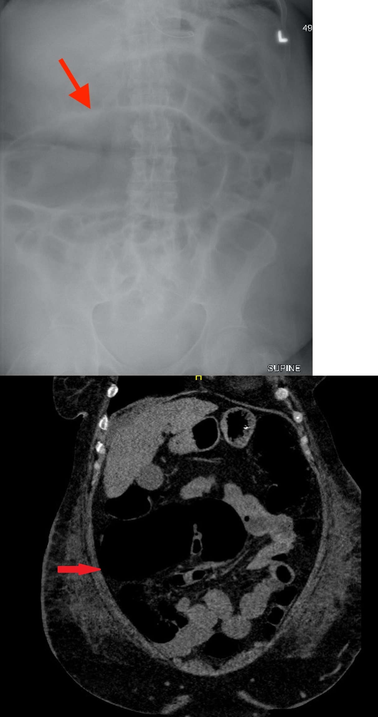
Case Description/Methods: A 63-year-old female with history of alcoholic cirrhosis (MELD 20) and cesarean section presented to the emergency department with intermittent hematochezia and melena. At admission, she was hemodynamically stable with a hemoglobin of 5.7 g/dL. She underwent esophagogastroduodenoscopy (EGD) which demonstrated small esophageal varices without stigmata of a recent bleed, antral gastritis, and duodenitis. Due to a largely unrevealing EGD, she was scheduled to have a colonoscopy. However, the patient had difficulties tolerating the bowel prep due to increasing abdominal pain. An abdominal X-ray demonstrated a large gaseous lucency in the right mid abdomen below the level of the transverse colon which was concerning for cecal bascule. A computed tomography (CT) scan of the abdomen demonstrated a redundant cecum folding anteriorly with superior rotation into the upper right hemiabdomen without definite point of transition and diffuse small bowel dilation up to 4.3 cm. Findings were consistent with a cecal bascule. Patient underwent an exploratory laparotomy with right hemicolectomy. Patient had a prolonged, complicated post-operative course with multiorgan failure and finally died 30 days after the surgery.
Discussion: Cecal bascule involves the upward folding of the cecum as opposed to an axial twisting of the colon as seen in more common types of cecal volvulus. For this phenomenon to occur, a patient will often have a mobile and redundant cecum that causes the volvulus. This may occur as a congenital anomaly secondary to a failed fusion in development between the ascending colon mesentery and the posterior parietal peritoneum. Additionally, this may be acquired from abdominal adhesions, pregnancy or even after a colonoscopy. Like other types of volvuli, treatment of cecal bascule requires surgical detorsion to prevent further complications. It is therefore important that cecal bascule be identified early for proper surgical planning and appropriate intervention. In our case, the poor outcome was due to the underlying decompensated cirrhosis.

Disclosures:
Jonathan E. Selzman, DO1, Mahesh Gajendran, MD, MPH2, Eric Smith, MD3, Chandraprakash Umapathy, MD4. A0160 - Cecal Bascule: A Rare Cause of Large Bowel Obstruction, ACG 2022 Annual Scientific Meeting Abstracts. Charlotte, NC: American College of Gastroenterology.
1University of Texas Health Science Center , San Antonio, TX; 2UTHSCSA, San Antonio, TX; 3BSW- Round Rock, Round Rock, TX; 4University of Texas Health San Antonio, San Antonio, TX
Introduction: Cecal bascule is a rare form of cecal volvulus, which is characterized by an anterior and superiorly displaced cecum in turn causing large bowel obstruction. It accounts for 0.01% of adult large bowel obstructions. Here we present a case of cecal bascule in a cirrhotic patient.
Case Description/Methods: A 63-year-old female with history of alcoholic cirrhosis (MELD 20) and cesarean section presented to the emergency department with intermittent hematochezia and melena. At admission, she was hemodynamically stable with a hemoglobin of 5.7 g/dL. She underwent esophagogastroduodenoscopy (EGD) which demonstrated small esophageal varices without stigmata of a recent bleed, antral gastritis, and duodenitis. Due to a largely unrevealing EGD, she was scheduled to have a colonoscopy. However, the patient had difficulties tolerating the bowel prep due to increasing abdominal pain. An abdominal X-ray demonstrated a large gaseous lucency in the right mid abdomen below the level of the transverse colon which was concerning for cecal bascule. A computed tomography (CT) scan of the abdomen demonstrated a redundant cecum folding anteriorly with superior rotation into the upper right hemiabdomen without definite point of transition and diffuse small bowel dilation up to 4.3 cm. Findings were consistent with a cecal bascule. Patient underwent an exploratory laparotomy with right hemicolectomy. Patient had a prolonged, complicated post-operative course with multiorgan failure and finally died 30 days after the surgery.
Discussion: Cecal bascule involves the upward folding of the cecum as opposed to an axial twisting of the colon as seen in more common types of cecal volvulus. For this phenomenon to occur, a patient will often have a mobile and redundant cecum that causes the volvulus. This may occur as a congenital anomaly secondary to a failed fusion in development between the ascending colon mesentery and the posterior parietal peritoneum. Additionally, this may be acquired from abdominal adhesions, pregnancy or even after a colonoscopy. Like other types of volvuli, treatment of cecal bascule requires surgical detorsion to prevent further complications. It is therefore important that cecal bascule be identified early for proper surgical planning and appropriate intervention. In our case, the poor outcome was due to the underlying decompensated cirrhosis.

Figure: Abdominal X-ray demonstrating a large gaseous lucency in the right mid abdomen near the transverse colon
CT of the abdomen demonstrating an anteriorly and superiorly rotated cecum into the upper right hemiabdomen
CT of the abdomen demonstrating an anteriorly and superiorly rotated cecum into the upper right hemiabdomen
Disclosures:
Jonathan Selzman indicated no relevant financial relationships.
Mahesh Gajendran indicated no relevant financial relationships.
Eric Smith indicated no relevant financial relationships.
Chandraprakash Umapathy indicated no relevant financial relationships.
Jonathan E. Selzman, DO1, Mahesh Gajendran, MD, MPH2, Eric Smith, MD3, Chandraprakash Umapathy, MD4. A0160 - Cecal Bascule: A Rare Cause of Large Bowel Obstruction, ACG 2022 Annual Scientific Meeting Abstracts. Charlotte, NC: American College of Gastroenterology.
