Back


Poster Session D - Tuesday Morning
Category: Interventional Endoscopy
D0456 - Not All Submucosal Lesions Are Created Equal: A Report of a Rare Cause of Abdominal Pain
Tuesday, October 25, 2022
10:00 AM – 12:00 PM ET
Location: Crown Ballroom

Has Audio
- KK
Katherine Kendrick, MD
Prisma Health
Greenville, SC
Presenting Author(s)
Katherine Kendrick, MD1, Jesse Clanton, MD2, Kristen Adams, MD1, Veeral Oza, MD3
1Prisma Health, Greenville, SC; 2Prisma Health, Columbia, SC; 3University of South Carolina School of Medicine Greenville, Prisma Health, Greenville, SC
Introduction: Gastric submucosal lesions are a common finding during endoscopy. However, they rarely present as or cause any symptoms. We present an interesting and rare case of a gastric submucosal lesion causing symptoms
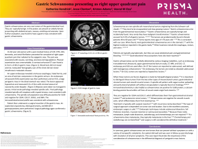
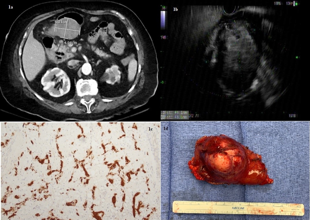
Case Description/Methods: An 82-year-old woman with multiple comorbidities presented with worsening nausea and abdominal pain and early satiety. Physical examination had no findings of abdominal tenderness or a palpable mass. A contrast enhanced computed tomography (CT) scan was then performed, and a 6.2cm x 4.9 cm gastric mass was noted. (Figure 1a)
An upper endoscopy revealed an extrinsic compression in the gastric antrum. An endoscopic ultrasound(EUS) examination was then performed, and on EUS, a large perigastric mass in the distal stomach was noted. The lesion appeared hypoechoic and measured 4.6 x 4.9cm with minimal amount of vascularity under EUS-doppler. (Fig1b) Transgastric biopsies were obtained. Final pathology revealed spindle cell neoplasm with myxoid stroma, most consistent with schwannoma. The immunohistochemical profile was consistent with schwannoma (Fig 1c). The patient then underwent surgical resection (Fig 1d) and pathology confirmed a gastric schwannoma
Discussion: Schwannomas are spindle cell mesenchymal tumors originating from the Schwann cell sheath. Gastric schwannomas arise from the gastrointestinal neural plexus. Gastric schwannomas are typically benign and incidentally found, but rarely can have malignant transformation. The tumors are predominantly found in middle-aged females. Gastric schwannoma usually presents in the gastric body.
It is important to distinguish these tumors from the two other types of mesenchymal tumors, such as gastrointestinal stromal tumors and leiomyoma. Schwannomas typically have a spindle cell pattern with vague nuclear palisading and peritumoral lymphoid cuff without encapsulation with S100 positive staining and negative for CD34 and CD117, which differentiates them from gastrointestinal stromal tumors and autonomic nerve tumors. Schwannomas stain negatively for actin, unlike leiomyomas.
Treatment is typically surgery with recurrence rarely described. The type of surgical approach is dependent on tumor size and location. Due to the excellent outcomes, some endoscopic options are also considered safe
Our patient did well and on follow had resolution of all prior symptoms. This case report illustrates the importance of the consideration of schwannomas in the differential diagnosis of perigastric and submucosal lesions

Disclosures:
Katherine Kendrick, MD1, Jesse Clanton, MD2, Kristen Adams, MD1, Veeral Oza, MD3. D0456 - Not All Submucosal Lesions Are Created Equal: A Report of a Rare Cause of Abdominal Pain, ACG 2022 Annual Scientific Meeting Abstracts. Charlotte, NC: American College of Gastroenterology.
1Prisma Health, Greenville, SC; 2Prisma Health, Columbia, SC; 3University of South Carolina School of Medicine Greenville, Prisma Health, Greenville, SC
Introduction: Gastric submucosal lesions are a common finding during endoscopy. However, they rarely present as or cause any symptoms. We present an interesting and rare case of a gastric submucosal lesion causing symptoms
Case Description/Methods: An 82-year-old woman with multiple comorbidities presented with worsening nausea and abdominal pain and early satiety. Physical examination had no findings of abdominal tenderness or a palpable mass. A contrast enhanced computed tomography (CT) scan was then performed, and a 6.2cm x 4.9 cm gastric mass was noted. (Figure 1a)
An upper endoscopy revealed an extrinsic compression in the gastric antrum. An endoscopic ultrasound(EUS) examination was then performed, and on EUS, a large perigastric mass in the distal stomach was noted. The lesion appeared hypoechoic and measured 4.6 x 4.9cm with minimal amount of vascularity under EUS-doppler. (Fig1b) Transgastric biopsies were obtained. Final pathology revealed spindle cell neoplasm with myxoid stroma, most consistent with schwannoma. The immunohistochemical profile was consistent with schwannoma (Fig 1c). The patient then underwent surgical resection (Fig 1d) and pathology confirmed a gastric schwannoma
Discussion: Schwannomas are spindle cell mesenchymal tumors originating from the Schwann cell sheath. Gastric schwannomas arise from the gastrointestinal neural plexus. Gastric schwannomas are typically benign and incidentally found, but rarely can have malignant transformation. The tumors are predominantly found in middle-aged females. Gastric schwannoma usually presents in the gastric body.
It is important to distinguish these tumors from the two other types of mesenchymal tumors, such as gastrointestinal stromal tumors and leiomyoma. Schwannomas typically have a spindle cell pattern with vague nuclear palisading and peritumoral lymphoid cuff without encapsulation with S100 positive staining and negative for CD34 and CD117, which differentiates them from gastrointestinal stromal tumors and autonomic nerve tumors. Schwannomas stain negatively for actin, unlike leiomyomas.
Treatment is typically surgery with recurrence rarely described. The type of surgical approach is dependent on tumor size and location. Due to the excellent outcomes, some endoscopic options are also considered safe
Our patient did well and on follow had resolution of all prior symptoms. This case report illustrates the importance of the consideration of schwannomas in the differential diagnosis of perigastric and submucosal lesions

Figure: Fig 1a: CT scan appearance of the perigastric mass; 1b: Mass as seen on EUS; 1c: Pathology with S100 staining; 1d: Resected specimen
Disclosures:
Katherine Kendrick indicated no relevant financial relationships.
Jesse Clanton indicated no relevant financial relationships.
Kristen Adams indicated no relevant financial relationships.
Veeral Oza: Boston Scientific – Consultant. S4 Medical – Advisor or Review Panel Member.
Katherine Kendrick, MD1, Jesse Clanton, MD2, Kristen Adams, MD1, Veeral Oza, MD3. D0456 - Not All Submucosal Lesions Are Created Equal: A Report of a Rare Cause of Abdominal Pain, ACG 2022 Annual Scientific Meeting Abstracts. Charlotte, NC: American College of Gastroenterology.
