Back


Poster Session D - Tuesday Morning
Category: Small Intestine
D0652 - Neuroendocrine Tumor of Ampulla of Vater in Existing Tubulovillous Adenoma
Tuesday, October 25, 2022
10:00 AM – 12:00 PM ET
Location: Crown Ballroom

Has Audio

Justine Chinnappan, MD
Hurley Medical Center
Flint, MI
Presenting Author(s)
Justine M. Chinnappan, MD1, Murtaza Hussain, MD2, Smit Deliwala, MD3, Ghassan Bachuwa, MD, MS, MHSA1, Qazi S. Azher, MD1, Mamoon Elbedawi, MD1
1Hurley Medical Center, Flint, MI; 2Hurley Medical Center at Michigan State University, Flint, MI; 3Emory University School of Medicine, Atlanta, GA
Introduction: Primary neuroendocrine carcinoma (NEC) of the ampulla of vater(AoV) is an extremely rare occurrence comprising 2% of periampullary neoplasms and < 1 % of gastrointestinal NEC. Likewise, ampullary adenoma is an uncommon occurrence with high predisposition for transformation to adenocarcinoma. Accordingly, their co-existence is sparsely reported. Herein we present a rare case of ampullary high grade neuroendocrine carcinoma (HGNEC) arising from a pre-existing tubulovillous adenoma.
Case Description/Methods: A 89 year old Jehovah witness male was evaluated for anemia 4 years ago and was found to have biopsy proven AoV tubulovillous adenoma. He was lost to follow-up after 6 months of surveillance esophagoduodenoscopy (EGD). He currently presents with severe bleeding per rectum with clots for one day. On presentation, he was hypotensive and with bright red blood mixed with stool on digital rectal exam. Laboratory work up revealed hemoglobin 8.1g/dL, alkaline phosphatase 748U/L, ALT 54U/L, AST 49U/L, Total bilirubin 1.8mg/dL and direct bilirubin 1.5mg/dL with elevated PT 17.6seconds. Given his ongoing bleeding, low hemoglobin and being a Jehovah witness, he was treated with IV iron sucrose, Aranesp, vitamin K, tranexamic acid. CT of the abdomen revealed extensive intra and extrahepatic bile duct dilation, common bile duct dilation (2.5 cm) and pancreatic duct dilation (1.2 cm). On MRCP, a small T2 hypointense lesion was identified near AoV. ERCP showed a large ulcerated mass measuring about 3 cm at the papilla, and the biopsy was significant for high grade small cell neuroendocrine carcinoma arising in a tubulovillous adenoma with immunohistochemical stains positive for CDX2, CD 56, SATB2 and synaptophysin. Ki-67 stain(cell proliferation marker) revealed positive in 100% of neuroendocrine cells. Biliary drain placement for hyperbilirubinemia (direct bilirubin 9.8mg/dL, 1 month from presentation) was done. Further course of action to be determined after correction of anemia and bilirubin.
Discussion: One study reported 50% of ampullary HGNEC is associated with adenoma but pathogenesis is still unclear. Although a HGNEC was identified in pre-existing adenoma, we need to further evaluate the possibility of adenocarcinoma arising from adenoma as this could affect further management of chemotherapy. The ampullary HGNEC identified appears aggressive with rapid growth and high Ki-67 index. Pancreatoduodenectomy with local lymph node dissection is warranted for concerns of early nodal metastasis at this time.

Disclosures:
Justine M. Chinnappan, MD1, Murtaza Hussain, MD2, Smit Deliwala, MD3, Ghassan Bachuwa, MD, MS, MHSA1, Qazi S. Azher, MD1, Mamoon Elbedawi, MD1. D0652 - Neuroendocrine Tumor of Ampulla of Vater in Existing Tubulovillous Adenoma, ACG 2022 Annual Scientific Meeting Abstracts. Charlotte, NC: American College of Gastroenterology.
1Hurley Medical Center, Flint, MI; 2Hurley Medical Center at Michigan State University, Flint, MI; 3Emory University School of Medicine, Atlanta, GA
Introduction: Primary neuroendocrine carcinoma (NEC) of the ampulla of vater(AoV) is an extremely rare occurrence comprising 2% of periampullary neoplasms and < 1 % of gastrointestinal NEC. Likewise, ampullary adenoma is an uncommon occurrence with high predisposition for transformation to adenocarcinoma. Accordingly, their co-existence is sparsely reported. Herein we present a rare case of ampullary high grade neuroendocrine carcinoma (HGNEC) arising from a pre-existing tubulovillous adenoma.
Case Description/Methods: A 89 year old Jehovah witness male was evaluated for anemia 4 years ago and was found to have biopsy proven AoV tubulovillous adenoma. He was lost to follow-up after 6 months of surveillance esophagoduodenoscopy (EGD). He currently presents with severe bleeding per rectum with clots for one day. On presentation, he was hypotensive and with bright red blood mixed with stool on digital rectal exam. Laboratory work up revealed hemoglobin 8.1g/dL, alkaline phosphatase 748U/L, ALT 54U/L, AST 49U/L, Total bilirubin 1.8mg/dL and direct bilirubin 1.5mg/dL with elevated PT 17.6seconds. Given his ongoing bleeding, low hemoglobin and being a Jehovah witness, he was treated with IV iron sucrose, Aranesp, vitamin K, tranexamic acid. CT of the abdomen revealed extensive intra and extrahepatic bile duct dilation, common bile duct dilation (2.5 cm) and pancreatic duct dilation (1.2 cm). On MRCP, a small T2 hypointense lesion was identified near AoV. ERCP showed a large ulcerated mass measuring about 3 cm at the papilla, and the biopsy was significant for high grade small cell neuroendocrine carcinoma arising in a tubulovillous adenoma with immunohistochemical stains positive for CDX2, CD 56, SATB2 and synaptophysin. Ki-67 stain(cell proliferation marker) revealed positive in 100% of neuroendocrine cells. Biliary drain placement for hyperbilirubinemia (direct bilirubin 9.8mg/dL, 1 month from presentation) was done. Further course of action to be determined after correction of anemia and bilirubin.
Discussion: One study reported 50% of ampullary HGNEC is associated with adenoma but pathogenesis is still unclear. Although a HGNEC was identified in pre-existing adenoma, we need to further evaluate the possibility of adenocarcinoma arising from adenoma as this could affect further management of chemotherapy. The ampullary HGNEC identified appears aggressive with rapid growth and high Ki-67 index. Pancreatoduodenectomy with local lymph node dissection is warranted for concerns of early nodal metastasis at this time.

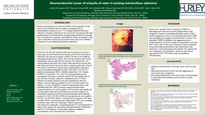
Figure: Figure 1: (A) Endoscopic image of large ulcerated ampullary tumor with bleeding (B) H & E stain X40 showing tubulovillous adenoma (black arrow) segment of normal AoV epithelium (blue arrow head) and nest of neuroendocrine cell (red arrow) (C) Immunohistochemical stain with synaptophysin x40 showing uniform staining of the neuroendocrine cells (red arrow)
Disclosures:
Justine Chinnappan indicated no relevant financial relationships.
Murtaza Hussain indicated no relevant financial relationships.
Smit Deliwala indicated no relevant financial relationships.
Ghassan Bachuwa indicated no relevant financial relationships.
Qazi Azher indicated no relevant financial relationships.
Mamoon Elbedawi indicated no relevant financial relationships.
Justine M. Chinnappan, MD1, Murtaza Hussain, MD2, Smit Deliwala, MD3, Ghassan Bachuwa, MD, MS, MHSA1, Qazi S. Azher, MD1, Mamoon Elbedawi, MD1. D0652 - Neuroendocrine Tumor of Ampulla of Vater in Existing Tubulovillous Adenoma, ACG 2022 Annual Scientific Meeting Abstracts. Charlotte, NC: American College of Gastroenterology.
