Back


Poster Session D - Tuesday Morning
Category: Small Intestine
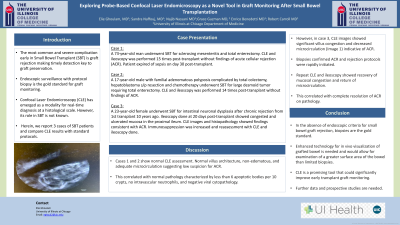
D0651 - Exploring Probe-Based Confocal Laser Endomicroscopy as a Novel Tool in Graft Monitoring After Small Bowel Transplantation
Tuesday, October 25, 2022
10:00 AM – 12:00 PM ET
Location: Crown Ballroom

Has Audio
- EG
Elie Ghoulam, MD
University of Ilinois at Chicago
Chicago, IL
Presenting Author(s)
Award: Presidential Poster Award
Elie Ghoulam, MD1, Sandra Naffouj, MD2, Najib Nassani, MD3, Grace Guzman, MD4, Enrico Benedetti, MD2, Robert E. Carroll, MD2
1University of Ilinois at Chicago, Chicago, IL; 2University of Illinois at Chicago, Chicago, IL; 3University of Chicago, Chicago, IL; 4University of Illinois Medical Center, Chicago, IL
Introduction: The most common and severe complication early in Small Bowel Transplant (SBT) is graft rejection making timely detection key to graft preservation. Endoscopic surveillance with protocol biopsy is the gold standard for graft monitoring. Confocal Laser Endomicroscopy (CLE) has emerged as a modality for real-time diagnosis at a histological scale. However, its role in SBT is not known. Herein, we report 3 cases of SBT patients and compare CLE results with standard protocols.
Case Description/Methods: Case 1:
A 73-year-old man underwent SBT for sclerosing mesenteritis and total enterectomy. CLE and ileoscopy was performed 15 times post-transplant without findings of acute cellular rejection (ACR). Patient expired of sepsis on day 30 post-transplant.
Case 2:
A 17-year-old male with familial adenomatous polyposis complicated by total colectomy, hepatoblastoma s/p resection and chemotherapy underwent SBT for large desmoid tumor requiring total enterectomy. CLE and ileoscopy was performed 14 times post-transplant without findings of ACR.
Case 3:
A 23-year-old female underwent SBT for intestinal neuronal dysplasia after chronic rejection from 1st transplant 10 years ago. Ileoscopy done at 20 days post-transplant showed congested and ulcerated mucosa in the proximal ileum. CLE images and histopathology showed findings consistent with ACR. Immunosuppression was increased and reassessment with CLE and ileoscopy done.
Discussion: Cases 1 and 2 show normal CLE assessment. Normal villus architecture, non-edematous, and adequate microcirculation suggesting low suspicion for ACR. This correlated with normal pathology characterized by less than 6 apoptotic bodies per 10 crypts, no intravascular neutrophils, and negative viral cytopathology. However, in case 3, CLE images showed significant villus congestion and decreased microcirculation (Image 1) indicative of ACR. Biopsies confirmed ACR. Rejection protocols were rapidly initiated.Repeat CLE and ileoscopy showed recovery of mucosal congestion and return of microcirculation. This correlated with complete resolution of ACR on pathology.
In the absence of endoscopic criteria for small bowel graft rejection, biopsies are the gold standard. Enhanced technology for in vivo visualization of grafted bowel is needed and would allow for examination of a greater surface area of the bowel than limited biopsies. CLE is a promising tool that could significantly improve early transplant graft monitoring. Further data and prospective studies are needed.

Disclosures:
Elie Ghoulam, MD1, Sandra Naffouj, MD2, Najib Nassani, MD3, Grace Guzman, MD4, Enrico Benedetti, MD2, Robert E. Carroll, MD2. D0651 - Exploring Probe-Based Confocal Laser Endomicroscopy as a Novel Tool in Graft Monitoring After Small Bowel Transplantation, ACG 2022 Annual Scientific Meeting Abstracts. Charlotte, NC: American College of Gastroenterology.
Elie Ghoulam, MD1, Sandra Naffouj, MD2, Najib Nassani, MD3, Grace Guzman, MD4, Enrico Benedetti, MD2, Robert E. Carroll, MD2
1University of Ilinois at Chicago, Chicago, IL; 2University of Illinois at Chicago, Chicago, IL; 3University of Chicago, Chicago, IL; 4University of Illinois Medical Center, Chicago, IL
Introduction: The most common and severe complication early in Small Bowel Transplant (SBT) is graft rejection making timely detection key to graft preservation. Endoscopic surveillance with protocol biopsy is the gold standard for graft monitoring. Confocal Laser Endomicroscopy (CLE) has emerged as a modality for real-time diagnosis at a histological scale. However, its role in SBT is not known. Herein, we report 3 cases of SBT patients and compare CLE results with standard protocols.
Case Description/Methods: Case 1:
A 73-year-old man underwent SBT for sclerosing mesenteritis and total enterectomy. CLE and ileoscopy was performed 15 times post-transplant without findings of acute cellular rejection (ACR). Patient expired of sepsis on day 30 post-transplant.
Case 2:
A 17-year-old male with familial adenomatous polyposis complicated by total colectomy, hepatoblastoma s/p resection and chemotherapy underwent SBT for large desmoid tumor requiring total enterectomy. CLE and ileoscopy was performed 14 times post-transplant without findings of ACR.
Case 3:
A 23-year-old female underwent SBT for intestinal neuronal dysplasia after chronic rejection from 1st transplant 10 years ago. Ileoscopy done at 20 days post-transplant showed congested and ulcerated mucosa in the proximal ileum. CLE images and histopathology showed findings consistent with ACR. Immunosuppression was increased and reassessment with CLE and ileoscopy done.
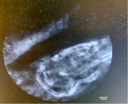
Discussion: Cases 1 and 2 show normal CLE assessment. Normal villus architecture, non-edematous, and adequate microcirculation suggesting low suspicion for ACR. This correlated with normal pathology characterized by less than 6 apoptotic bodies per 10 crypts, no intravascular neutrophils, and negative viral cytopathology. However, in case 3, CLE images showed significant villus congestion and decreased microcirculation (Image 1) indicative of ACR. Biopsies confirmed ACR. Rejection protocols were rapidly initiated.Repeat CLE and ileoscopy showed recovery of mucosal congestion and return of microcirculation. This correlated with complete resolution of ACR on pathology.
In the absence of endoscopic criteria for small bowel graft rejection, biopsies are the gold standard. Enhanced technology for in vivo visualization of grafted bowel is needed and would allow for examination of a greater surface area of the bowel than limited biopsies. CLE is a promising tool that could significantly improve early transplant graft monitoring. Further data and prospective studies are needed.

Figure: CLE images showing significant villus congestion, and decreased microcirculation
Disclosures:
Elie Ghoulam indicated no relevant financial relationships.
Sandra Naffouj indicated no relevant financial relationships.
Najib Nassani indicated no relevant financial relationships.
Grace Guzman indicated no relevant financial relationships.
Enrico Benedetti indicated no relevant financial relationships.
Robert Carroll indicated no relevant financial relationships.
Elie Ghoulam, MD1, Sandra Naffouj, MD2, Najib Nassani, MD3, Grace Guzman, MD4, Enrico Benedetti, MD2, Robert E. Carroll, MD2. D0651 - Exploring Probe-Based Confocal Laser Endomicroscopy as a Novel Tool in Graft Monitoring After Small Bowel Transplantation, ACG 2022 Annual Scientific Meeting Abstracts. Charlotte, NC: American College of Gastroenterology.

