Back


Poster Session D - Tuesday Morning
Category: Stomach
D0709 - Metastatic Gastric Adenocarcinoma in 23-Year-Old Female
Tuesday, October 25, 2022
10:00 AM – 12:00 PM ET
Location: Crown Ballroom

Has Audio

Khandokar A. Talib, MD
United Health Services Hospital
Johnson City, NY
Presenting Author(s)
Khandokar A. Talib, MD, Gowthami Ramar, MD, Usama Shakhwat, MD, Khaleeq Siddiqui, MD, Minhaz Ahmad, MD, Mark Shumeyko, MD
United Health Services Hospital, Johnson City, NY
Introduction: Gastric cancer is the fourth most common malignancy in the USA. Since there are no screening recommendations in place, most gastric cancers are detected at a later stage in the USA. The average age of occurrence of gastric cancer is >40 years. Some of the most common risk factors include Helicobacter pylori infection, atrophic gastritis, ethnicity, and dietary factors.
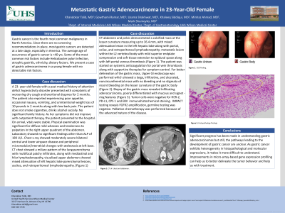
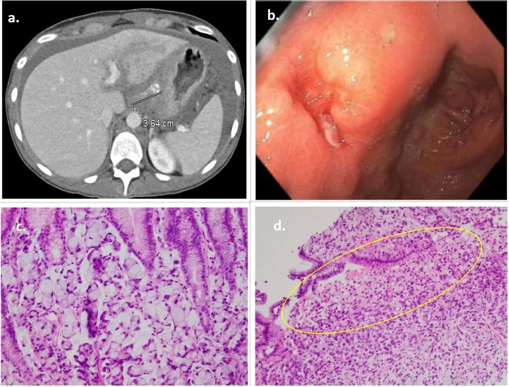
Case Description/Methods: A 23-year-old female with a past medical history of attention deficit hyperactivity disorder presented with complaints of worsening cough and exertional dyspnea for 2 months. The patient also reported experiencing poor appetite, occasional nausea, vomiting, and unintentional weight loss of 10 pounds in 2 months with low back pain. The patient does not smoke cigarettes and drinks alcohol socially. No significant family history. Physical examination was unremarkable. Labs were normal except for mildly elevated ALP. Chest x-ray showed bilateral infiltrates. CT chest showed a miliary pattern of the lung parenchyma with multifocal patchy infiltrates and mediastinal and hilar lymphadenopathy. CT abdomen and pelvis (Figure 1) showed a calcified mass at the lesser curvature measuring up to 36.4 mm, with mixed attenuation lesion in the left hepatic lobe along with portal, celiac, and retroperitoneal lymphadenopathy; metastatic lesion within the L2 vertebral body with mild superior endplate compression and soft tissue extension to epidural space along with left portal venous thrombosis. Upper GI endoscopy (Figure 1) showed a large, infiltrative, ulcerated, noncircumferential mass on the lower curvature of the gastric body. Biopsy of the gastric mass revealed infiltrative adenocarcinoma, poorly differentiated with mucous and signet-ring features. Tumor cells were negative for E-cadherin expression and HER-2 immunohistochemical staining and positive for PD-L1 and MYC amplification. The plan was to perform palliative chemotherapy because of the advanced nature of the disease.
Discussion: Significant progress has been made in understanding gastric adenocarcinomas, but the pathways leading to the development of gastric cancer are still unclear. As gastric cancer exhibits heterogeneity and molecular expressions, it makes it more difficult to understand. Improvements in micro-array-based gene expression profiling can help us to better delineate the tumor behavior and help us with treatment.

Disclosures:
Khandokar A. Talib, MD, Gowthami Ramar, MD, Usama Shakhwat, MD, Khaleeq Siddiqui, MD, Minhaz Ahmad, MD, Mark Shumeyko, MD. D0709 - Metastatic Gastric Adenocarcinoma in 23-Year-Old Female, ACG 2022 Annual Scientific Meeting Abstracts. Charlotte, NC: American College of Gastroenterology.
United Health Services Hospital, Johnson City, NY
Introduction: Gastric cancer is the fourth most common malignancy in the USA. Since there are no screening recommendations in place, most gastric cancers are detected at a later stage in the USA. The average age of occurrence of gastric cancer is >40 years. Some of the most common risk factors include Helicobacter pylori infection, atrophic gastritis, ethnicity, and dietary factors.
Case Description/Methods: A 23-year-old female with a past medical history of attention deficit hyperactivity disorder presented with complaints of worsening cough and exertional dyspnea for 2 months. The patient also reported experiencing poor appetite, occasional nausea, vomiting, and unintentional weight loss of 10 pounds in 2 months with low back pain. The patient does not smoke cigarettes and drinks alcohol socially. No significant family history. Physical examination was unremarkable. Labs were normal except for mildly elevated ALP. Chest x-ray showed bilateral infiltrates. CT chest showed a miliary pattern of the lung parenchyma with multifocal patchy infiltrates and mediastinal and hilar lymphadenopathy. CT abdomen and pelvis (Figure 1) showed a calcified mass at the lesser curvature measuring up to 36.4 mm, with mixed attenuation lesion in the left hepatic lobe along with portal, celiac, and retroperitoneal lymphadenopathy; metastatic lesion within the L2 vertebral body with mild superior endplate compression and soft tissue extension to epidural space along with left portal venous thrombosis. Upper GI endoscopy (Figure 1) showed a large, infiltrative, ulcerated, noncircumferential mass on the lower curvature of the gastric body. Biopsy of the gastric mass revealed infiltrative adenocarcinoma, poorly differentiated with mucous and signet-ring features. Tumor cells were negative for E-cadherin expression and HER-2 immunohistochemical staining and positive for PD-L1 and MYC amplification. The plan was to perform palliative chemotherapy because of the advanced nature of the disease.
Discussion: Significant progress has been made in understanding gastric adenocarcinomas, but the pathways leading to the development of gastric cancer are still unclear. As gastric cancer exhibits heterogeneity and molecular expressions, it makes it more difficult to understand. Improvements in micro-array-based gene expression profiling can help us to better delineate the tumor behavior and help us with treatment.

Figure: Figure 1: a. CT abdomen - partially calcified gastric mass; b. EGD - A large infiltrative and ulcerated, noncircumferential mass on the lesser curvature of the gastric body; c. & d. Histopathology - H&E stain (20 x): gastric mucosa infiltrated by carcinoma cells with a signet-ring morphology.
Disclosures:
Khandokar Talib indicated no relevant financial relationships.
Gowthami Ramar indicated no relevant financial relationships.
Usama Shakhwat indicated no relevant financial relationships.
Khaleeq Siddiqui indicated no relevant financial relationships.
Minhaz Ahmad indicated no relevant financial relationships.
Mark Shumeyko indicated no relevant financial relationships.
Khandokar A. Talib, MD, Gowthami Ramar, MD, Usama Shakhwat, MD, Khaleeq Siddiqui, MD, Minhaz Ahmad, MD, Mark Shumeyko, MD. D0709 - Metastatic Gastric Adenocarcinoma in 23-Year-Old Female, ACG 2022 Annual Scientific Meeting Abstracts. Charlotte, NC: American College of Gastroenterology.
