Back


Poster Session D - Tuesday Morning
Category: Small Intestine
D0679 - Duodenal Mass With Lymphadenopathy: A Case of Gastrointestinal Coccidioidomycosis
Tuesday, October 25, 2022
10:00 AM – 12:00 PM ET
Location: Crown Ballroom

Has Audio

Idrees Suliman, MD
Mountain Vista Medical Center
Mesa, AZ
Presenting Author(s)
Idrees Suliman, MD1, Lyndie R. Wilkins Parker, DO1, Raxitkumar Patel, MD2, Paresh Sojitra, MD2, Sudhakar Reddy, MD2, Preeyanka Sundar, MD2, Abdul Nadir, MD1
1Mountain Vista Medical Center, Mesa, AZ; 2Midwestern University, Mesa, AZ
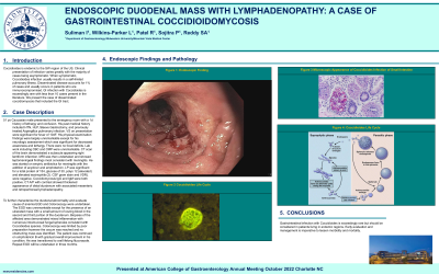
Introduction: Coccidioides is endemic to the SW region of the US. Clinical presentation of infection varies greatly with the majority of cases being asymptomatic. When symptomatic, Coccidioides infection usually results in a self-limited pulmonary illness. Disseminated disease accounts for 1% of cases and usually occurs in patients who are immunocompromised. GI infection with Coccidioides is exceedingly rare with less than 10 cases present in the literature. We present the case of disseminated coccidiomycosis that included the GI tract.
Case Description/Methods: 51 yo Caucasian male presented to the emergency room with a 1d history of lethargy and
confusion. His past medical history included HTN, HLP, RnY gastric bypass, and
previously treated Aspergillus pulmonary infection. VS on presentation were significant for fever of 104
F. His physical examination findings were largely unremarkable except for his neurologic assessment which
was significant for decreased awareness and lethargy. There were no focal deficits. Lab work including CBC
and CMP were unremarkable. CT scan of the brain demonstrated a subacute appearing right lentiform
infarction. MRI was then undertaken and showed leptomeningeal findings most consistent with meningitis. He
was started on empiric antibiotics for meningitis with the addition of acyclovir and amphotericin. LP was significant for a total protein of 154, glucose of 30, polys 12 (elevated) and elevated eosinophils
(2). CSF gram stain and VDRL were negative. Coccidiomycosis IgG and IgM were both positive. CT A/P with
contrast showed thickened appearance of distal duodenum with associated mesenteric and retroperitoneal
lymphadenopathy.
To further characterize the duodenal abnormality and evaluate cause of anemia EGD and Colonoscopy were
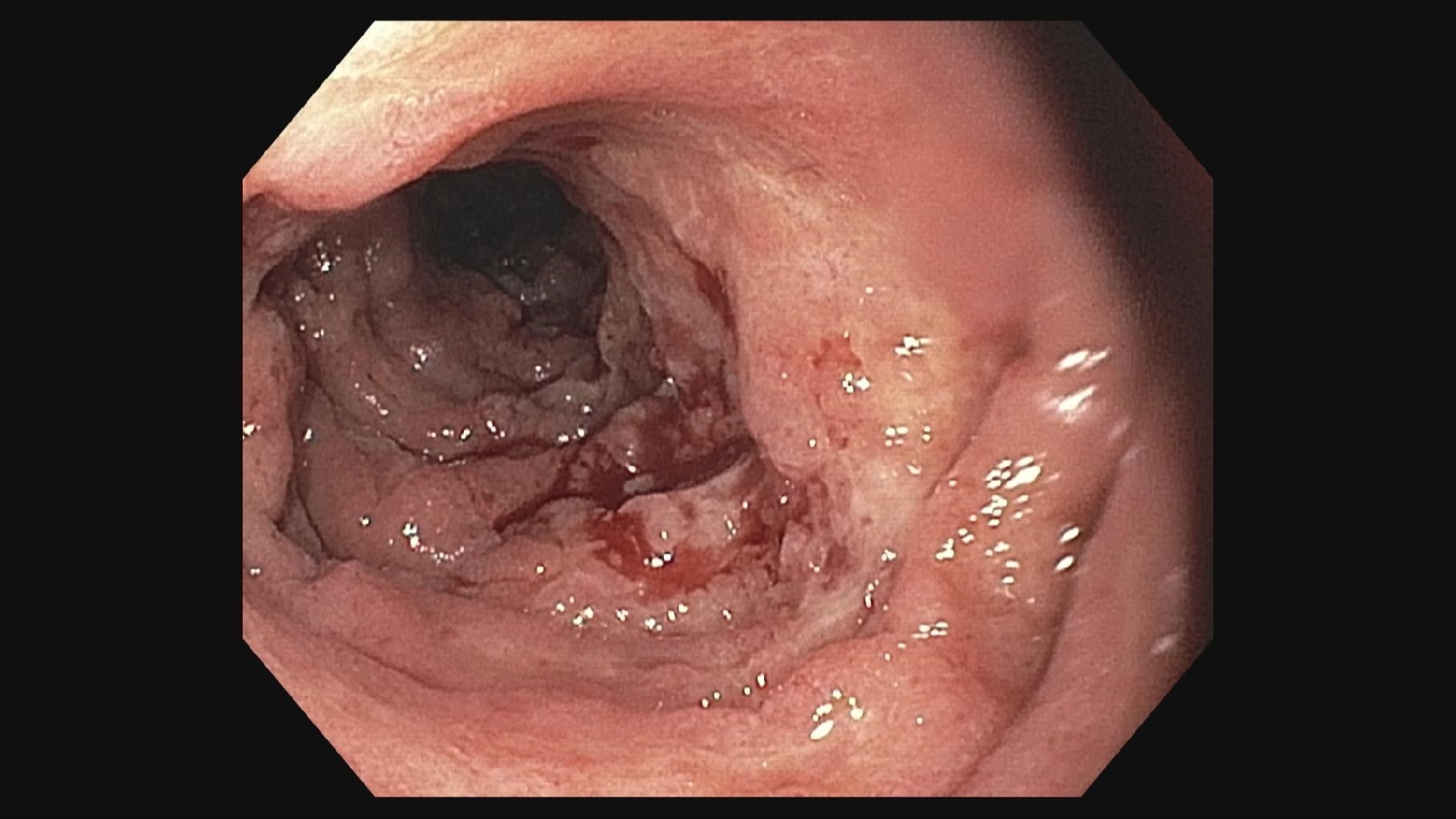
undertaken. The EGD was unremarkable except for the presence of an ulcerated mass with a small amount of
oozing blood in the second and third portion of the duodenum. Biopsies of the affected area demonstrated
mixed inflammation with numerous intramucosal fungal spherules consistent with Coccidioides species.
Colonoscopy was limited by poor preparation however the cecum was reached and no obstructing mass was
identified. The patient was continued on amphotericin B with gradual overall improvement in his condition. He
was transitioned to oral lifelong fluconazole. Repeat EGD will be undertaken in three months.
Discussion: Gastrointestinal infection with Coccidioides is exceedingly rare but should be considered in patients living in endemic regions.

Disclosures:
Idrees Suliman, MD1, Lyndie R. Wilkins Parker, DO1, Raxitkumar Patel, MD2, Paresh Sojitra, MD2, Sudhakar Reddy, MD2, Preeyanka Sundar, MD2, Abdul Nadir, MD1. D0679 - Duodenal Mass With Lymphadenopathy: A Case of Gastrointestinal Coccidioidomycosis, ACG 2022 Annual Scientific Meeting Abstracts. Charlotte, NC: American College of Gastroenterology.
1Mountain Vista Medical Center, Mesa, AZ; 2Midwestern University, Mesa, AZ
Introduction: Coccidioides is endemic to the SW region of the US. Clinical presentation of infection varies greatly with the majority of cases being asymptomatic. When symptomatic, Coccidioides infection usually results in a self-limited pulmonary illness. Disseminated disease accounts for 1% of cases and usually occurs in patients who are immunocompromised. GI infection with Coccidioides is exceedingly rare with less than 10 cases present in the literature. We present the case of disseminated coccidiomycosis that included the GI tract.
Case Description/Methods: 51 yo Caucasian male presented to the emergency room with a 1d history of lethargy and
confusion. His past medical history included HTN, HLP, RnY gastric bypass, and
previously treated Aspergillus pulmonary infection. VS on presentation were significant for fever of 104
F. His physical examination findings were largely unremarkable except for his neurologic assessment which
was significant for decreased awareness and lethargy. There were no focal deficits. Lab work including CBC
and CMP were unremarkable. CT scan of the brain demonstrated a subacute appearing right lentiform
infarction. MRI was then undertaken and showed leptomeningeal findings most consistent with meningitis. He
was started on empiric antibiotics for meningitis with the addition of acyclovir and amphotericin. LP was significant for a total protein of 154, glucose of 30, polys 12 (elevated) and elevated eosinophils
(2). CSF gram stain and VDRL were negative. Coccidiomycosis IgG and IgM were both positive. CT A/P with
contrast showed thickened appearance of distal duodenum with associated mesenteric and retroperitoneal
lymphadenopathy.
To further characterize the duodenal abnormality and evaluate cause of anemia EGD and Colonoscopy were
undertaken. The EGD was unremarkable except for the presence of an ulcerated mass with a small amount of
oozing blood in the second and third portion of the duodenum. Biopsies of the affected area demonstrated
mixed inflammation with numerous intramucosal fungal spherules consistent with Coccidioides species.
Colonoscopy was limited by poor preparation however the cecum was reached and no obstructing mass was
identified. The patient was continued on amphotericin B with gradual overall improvement in his condition. He
was transitioned to oral lifelong fluconazole. Repeat EGD will be undertaken in three months.
Discussion: Gastrointestinal infection with Coccidioides is exceedingly rare but should be considered in patients living in endemic regions.

Figure: Duodenum
Disclosures:
Idrees Suliman indicated no relevant financial relationships.
Lyndie Wilkins Parker indicated no relevant financial relationships.
Raxitkumar Patel indicated no relevant financial relationships.
Paresh Sojitra indicated no relevant financial relationships.
Sudhakar Reddy indicated no relevant financial relationships.
Preeyanka Sundar indicated no relevant financial relationships.
Abdul Nadir indicated no relevant financial relationships.
Idrees Suliman, MD1, Lyndie R. Wilkins Parker, DO1, Raxitkumar Patel, MD2, Paresh Sojitra, MD2, Sudhakar Reddy, MD2, Preeyanka Sundar, MD2, Abdul Nadir, MD1. D0679 - Duodenal Mass With Lymphadenopathy: A Case of Gastrointestinal Coccidioidomycosis, ACG 2022 Annual Scientific Meeting Abstracts. Charlotte, NC: American College of Gastroenterology.
