Back


Poster Session E - Tuesday Afternoon
Category: Colon
E0138 - Ulcerative Rectal Mass Complicated by Recto Cutaneous Fistula: Cytomegalovirus as a Culprit
Tuesday, October 25, 2022
3:00 PM – 5:00 PM ET
Location: Crown Ballroom

Has Audio

Rameela Mahat, MD
Baton Rouge General Internal Medicine Residency Program
Baton Rouge, LA
Presenting Author(s)
Rameela Mahat, MD1, Jeremy Polman, DO, MS, MBA2, Aaron Williams, MD3
1Baton Rouge General Internal Medicine Residency Program, Baton Rouge, LA; 2Baton Rouge General Medical Center, Baton Rouge, LA; 3HMG Baton Rouge Clinic, Baton Rouge, LA
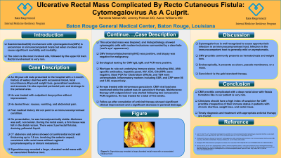
Introduction: Gastrointestinal(GI) involvement with cytomegalovirus(CMV) is uncommon in immunocompetent hosts. The colon is the most commonly involved site followed by the upper GI tract. Rectal involvement is very rare.
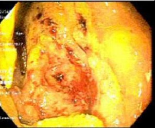
Case Description/Methods: An 80-year-old male presented to the hospital with a 3-month history of watery diarrhea with occasional blood, fecal incontinence,40-pound weight loss, generalized weakness, anorexia, perirectal pain, and drainage in the perianal area. He was treated with outpatient doxycycline without improvement. He denied fever, nausea, vomiting, and abdominal pain. Past medical history did not point to an immunocompromised condition. On presentation, he was hemodynamically stable. Abdomen was soft, non tender. During the rectal exam, a firm tissue was felt in the distal rectum. There were 3 perirectal fistulas, draining yellowish liquid. CT abdomen and pelvis showed circumferential rectal wall thickening up to 1.8 cm, involving anterior aspect consistent with rectal mass without regional lymphadenopathy or distant metastasis. Sigmoidoscopy revealed an ulcerated rectal mass with an associated fistulous tract, with negative biopsies for malignancy and positive immunohistochemistry(IHC) for CMV. Serological testing for CMV IgG, IgM, and PCR were positive. Workups including ANA, hepatitis panel, HIV RPR, Stool PCR for Clostridium difficile, and TSH were unremarkable. He was treated with Intravenous Ganciclovir. After two consecutive PCR negatives for CMV, maintenance therapy with Valganciclovir was started for a total of 5 weeks. He was followed in the outpatient setting. His symptoms were improved. He also had a significant decrease in perianal drainage.
Discussion: Cytomegalovirus is well recognized to cause opportunistic infection in an immunocompromised host. Infection in the immunocompetent host is generally mild or asymptomatic. However, in rare situations, it can lead to severe infection with substantial mortality and morbidity. CMV proctitis commonly presents as hematochezia, and weight loss. Endoscopically it presents as ulcers, pseudo membranes, or a mass. CMV proctitis complicated with a deep rectal ulcer with fistula formation like in our patient is very rare. Ganciclovir is the gold standard therapy. Clinicians should have a high index of suspicion for CMV proctitis irrespective of their immune status in patients with chronic diarrhea, weight loss, and rectal mass. Timely diagnosis and treatment with appropriate antiviral therapy are crucial.

Disclosures:
Rameela Mahat, MD1, Jeremy Polman, DO, MS, MBA2, Aaron Williams, MD3. E0138 - Ulcerative Rectal Mass Complicated by Recto Cutaneous Fistula: Cytomegalovirus as a Culprit, ACG 2022 Annual Scientific Meeting Abstracts. Charlotte, NC: American College of Gastroenterology.
1Baton Rouge General Internal Medicine Residency Program, Baton Rouge, LA; 2Baton Rouge General Medical Center, Baton Rouge, LA; 3HMG Baton Rouge Clinic, Baton Rouge, LA
Introduction: Gastrointestinal(GI) involvement with cytomegalovirus(CMV) is uncommon in immunocompetent hosts. The colon is the most commonly involved site followed by the upper GI tract. Rectal involvement is very rare.
Case Description/Methods: An 80-year-old male presented to the hospital with a 3-month history of watery diarrhea with occasional blood, fecal incontinence,40-pound weight loss, generalized weakness, anorexia, perirectal pain, and drainage in the perianal area. He was treated with outpatient doxycycline without improvement. He denied fever, nausea, vomiting, and abdominal pain. Past medical history did not point to an immunocompromised condition. On presentation, he was hemodynamically stable. Abdomen was soft, non tender. During the rectal exam, a firm tissue was felt in the distal rectum. There were 3 perirectal fistulas, draining yellowish liquid. CT abdomen and pelvis showed circumferential rectal wall thickening up to 1.8 cm, involving anterior aspect consistent with rectal mass without regional lymphadenopathy or distant metastasis. Sigmoidoscopy revealed an ulcerated rectal mass with an associated fistulous tract, with negative biopsies for malignancy and positive immunohistochemistry(IHC) for CMV. Serological testing for CMV IgG, IgM, and PCR were positive. Workups including ANA, hepatitis panel, HIV RPR, Stool PCR for Clostridium difficile, and TSH were unremarkable. He was treated with Intravenous Ganciclovir. After two consecutive PCR negatives for CMV, maintenance therapy with Valganciclovir was started for a total of 5 weeks. He was followed in the outpatient setting. His symptoms were improved. He also had a significant decrease in perianal drainage.
Discussion: Cytomegalovirus is well recognized to cause opportunistic infection in an immunocompromised host. Infection in the immunocompetent host is generally mild or asymptomatic. However, in rare situations, it can lead to severe infection with substantial mortality and morbidity. CMV proctitis commonly presents as hematochezia, and weight loss. Endoscopically it presents as ulcers, pseudo membranes, or a mass. CMV proctitis complicated with a deep rectal ulcer with fistula formation like in our patient is very rare. Ganciclovir is the gold standard therapy. Clinicians should have a high index of suspicion for CMV proctitis irrespective of their immune status in patients with chronic diarrhea, weight loss, and rectal mass. Timely diagnosis and treatment with appropriate antiviral therapy are crucial.

Figure: Flexible Sigmoidoscopy showing ulcerated rectal mass with the surrounding area of erythematous, furrowed rectal mucosa.
Disclosures:
Rameela Mahat indicated no relevant financial relationships.
Jeremy Polman indicated no relevant financial relationships.
Aaron Williams indicated no relevant financial relationships.
Rameela Mahat, MD1, Jeremy Polman, DO, MS, MBA2, Aaron Williams, MD3. E0138 - Ulcerative Rectal Mass Complicated by Recto Cutaneous Fistula: Cytomegalovirus as a Culprit, ACG 2022 Annual Scientific Meeting Abstracts. Charlotte, NC: American College of Gastroenterology.
