Back


Poster Session E - Tuesday Afternoon
Category: Colon
E0142 - Anorectal Mucosal Melanoma: A Case Series Demonstrating the Importance of Maintaining a High Index of Suspicion for This Deadly Disease
Tuesday, October 25, 2022
3:00 PM – 5:00 PM ET
Location: Crown Ballroom

Has Audio

Jeremy Polman, DO, MS, MBA
Baton Rouge General Medical Center
Baton Rouge, LA
Presenting Author(s)
Award: Presidential Poster Award
Jeremy Polman, DO, MS, MBA1, Gabrielle Sanford, MD2, Marshall Stagg, MD2, Rameela Mahat, MD3
1Baton Rouge General Medical Center, Baton Rouge, LA; 2LSUHSC Baton Rouge Internal Medicine Residency Program at Our Lady of the Lake, Baton Rouge, LA; 3Baton Rouge General Internal Medicine Residency Program, Baton Rouge, LA
Introduction: Anorectal Mucosal Melanoma (ARM) is a rare and aggressive malignancy that is poorly described and infrequently studied. ARM differs significantly from cutaneous melanoma in presentation, genetic profile, staging, treatment response, and progression patterns. ARM is a challenge for physicians because of its rarity, unclear pathogenesis, non-specific presentation, and aggressive disease course leading to an inferior prognosis. Consequently, it can be misdiagnosed clinically as a benign disease. This report presents three clinical cases, all of which presented with relatively benign symptoms, and outlines the extensive evaluation and treatment of these patients.
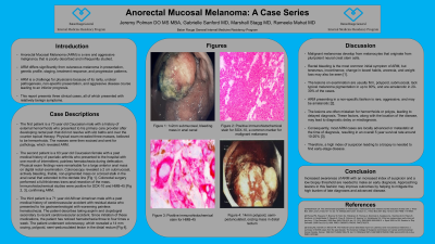
Case Description/Methods: The first patient is a 73 year old Caucasian male with a past medical history of external hemorrhoids who presented to his primary care provider after developing rectal pain that did not resolve with sitz baths and over the counter topical therapy. Physical exam revealed three masses, believed to be hemorrhoids. The masses were then excised and sent for pathology, which revealed ARM. The second patient is a 63 year old Caucasian female with a past medical history of psoriatic arthritis who presented to the hospital with one month of intermittent, painless hematochezia during defecation. Physical exam findings were remarkable for a large anterior anal mass on digital rectal examination. Colonoscopy revealed a 2 cm submucosal, actively bleeding, friable, non-pigmented mass on a broad stalk in the anal canal that extended to the dentate line [A]. Colorectal surgery performed a full-thickness trans-anal resection of the mass. Immunohistochemical studies were positive for SOX-10 and Melan-A [B, C], confirming ARM. The third patient is a 71 year old African American male with a past medical history of cerebrovascular accident with residual ataxia who presented to his gastroenterologist with worsening painless hematochezia. The patient describes taking aspirin and clopidogrel secondary to recent cerebrovascular accident. Since initiation of these medications, the patient has noticed hematochezia three to four times a week. The patient underwent colonoscopy, which revealed a 14 mm oozing, polypoid, semi-pedunculated lesion in the distal rectum.
Discussion: Increased awareness of ARM with an increased index of suspicion and a low biopsy threshold is needed to make an early diagnosis. Approaching lesions in this fashion may improve outcomes by helping to mitigate the high burden of late diagnoses and advanced disease.

Disclosures:
Jeremy Polman, DO, MS, MBA1, Gabrielle Sanford, MD2, Marshall Stagg, MD2, Rameela Mahat, MD3. E0142 - Anorectal Mucosal Melanoma: A Case Series Demonstrating the Importance of Maintaining a High Index of Suspicion for This Deadly Disease, ACG 2022 Annual Scientific Meeting Abstracts. Charlotte, NC: American College of Gastroenterology.
Jeremy Polman, DO, MS, MBA1, Gabrielle Sanford, MD2, Marshall Stagg, MD2, Rameela Mahat, MD3
1Baton Rouge General Medical Center, Baton Rouge, LA; 2LSUHSC Baton Rouge Internal Medicine Residency Program at Our Lady of the Lake, Baton Rouge, LA; 3Baton Rouge General Internal Medicine Residency Program, Baton Rouge, LA
Introduction: Anorectal Mucosal Melanoma (ARM) is a rare and aggressive malignancy that is poorly described and infrequently studied. ARM differs significantly from cutaneous melanoma in presentation, genetic profile, staging, treatment response, and progression patterns. ARM is a challenge for physicians because of its rarity, unclear pathogenesis, non-specific presentation, and aggressive disease course leading to an inferior prognosis. Consequently, it can be misdiagnosed clinically as a benign disease. This report presents three clinical cases, all of which presented with relatively benign symptoms, and outlines the extensive evaluation and treatment of these patients.
Case Description/Methods: The first patient is a 73 year old Caucasian male with a past medical history of external hemorrhoids who presented to his primary care provider after developing rectal pain that did not resolve with sitz baths and over the counter topical therapy. Physical exam revealed three masses, believed to be hemorrhoids. The masses were then excised and sent for pathology, which revealed ARM. The second patient is a 63 year old Caucasian female with a past medical history of psoriatic arthritis who presented to the hospital with one month of intermittent, painless hematochezia during defecation. Physical exam findings were remarkable for a large anterior anal mass on digital rectal examination. Colonoscopy revealed a 2 cm submucosal, actively bleeding, friable, non-pigmented mass on a broad stalk in the anal canal that extended to the dentate line [A]. Colorectal surgery performed a full-thickness trans-anal resection of the mass. Immunohistochemical studies were positive for SOX-10 and Melan-A [B, C], confirming ARM. The third patient is a 71 year old African American male with a past medical history of cerebrovascular accident with residual ataxia who presented to his gastroenterologist with worsening painless hematochezia. The patient describes taking aspirin and clopidogrel secondary to recent cerebrovascular accident. Since initiation of these medications, the patient has noticed hematochezia three to four times a week. The patient underwent colonoscopy, which revealed a 14 mm oozing, polypoid, semi-pedunculated lesion in the distal rectum.
Discussion: Increased awareness of ARM with an increased index of suspicion and a low biopsy threshold is needed to make an early diagnosis. Approaching lesions in this fashion may improve outcomes by helping to mitigate the high burden of late diagnoses and advanced disease.

Figure: A) Image obtained during colonoscopy showing 1x2 cm, submucosal, bleeding, friable, benign appearing, fibrovascular mass in the anal canal, extending distal to the dentate line.
B) Tumor biopsy demonstrating positive to immunohistochemical stain for SOX-10, a common marker for malignant melanoma.
C) Tumor biopsy with immunohistochemical stain for Melan-A positivity.
B) Tumor biopsy demonstrating positive to immunohistochemical stain for SOX-10, a common marker for malignant melanoma.
C) Tumor biopsy with immunohistochemical stain for Melan-A positivity.
Disclosures:
Jeremy Polman indicated no relevant financial relationships.
Gabrielle Sanford indicated no relevant financial relationships.
Marshall Stagg indicated no relevant financial relationships.
Rameela Mahat indicated no relevant financial relationships.
Jeremy Polman, DO, MS, MBA1, Gabrielle Sanford, MD2, Marshall Stagg, MD2, Rameela Mahat, MD3. E0142 - Anorectal Mucosal Melanoma: A Case Series Demonstrating the Importance of Maintaining a High Index of Suspicion for This Deadly Disease, ACG 2022 Annual Scientific Meeting Abstracts. Charlotte, NC: American College of Gastroenterology.

