Back


Poster Session D - Tuesday Morning
Category: Biliary/Pancreas
D0038 - An Atypical, Silent Spread of Gallbladder Cancer
Tuesday, October 25, 2022
10:00 AM – 12:00 PM ET
Location: Crown Ballroom

Has Audio

Rami Abusaleh, MD
Creighton University
Tempe, AZ
Presenting Author(s)
Rami Abusaleh, MD1, Chinonso Ilo, MD1, Aida Rezaie, MD1, Melinda Wang, MD1, Indu Srinivasan, MD2, Keng-Yu Chuang, MD2
1Creighton University, Phoenix, AZ; 2Valleywise Health, Phoenix, AZ
Introduction: Gallbladder cancer (GBC) though rare is the most common biliary cancer. It is associated with a poor prognosis due to delayed diagnosis and nonspecific clinical presentation. We present a case of a 62-year-old female that was incidentally found to have metastatic gallbladder cancer with no alarm symptoms on presentation.
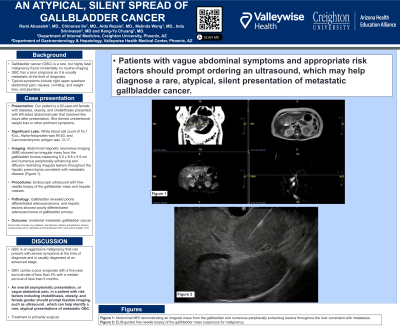
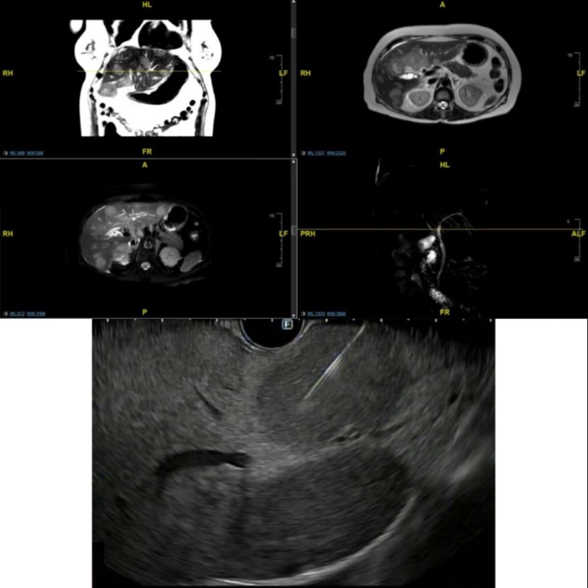
Case Description/Methods: A 62-year-old female with diabetes mellitus II, obesity, and cholelithiasis presented to the emergency room with vague complaints of lower abdominal pain. Lab work was grossly normal except elevated white blood cell count of 16.1 K/uL. The pain resolved with bowel movement, however initial CT scan obtained was concerning for gall bladder mass. Tumor markers were obtained. Alpha-fetoprotein was 94.63, carcinoembryonic antigen was 13.17, and cancer antigen 19-9 was 24 within normal limits. Abdominal Magnetic resonance imaging (MRI) done for further evaluation showed an irregular mass from the gallbladder fundus measuring 5.0 x 8.8 x 5.9 cm along with numerous peripherally enhancing and diffusion restricting irregular lesions throughout the hepatic parenchyma consistent with metastatic disease. The patient underwent endoscopic ultrasound with fine-needle biopsy of the lesions and pathology was consistent with poorly differentiated adenocarcinoma of gallbladder primary.
Discussion: Gallbladder cancer (GBC) is an aggressive malignancy that is usually diagnosed at an advanced stage and thus carries a poor prognosis with a five-year survival rate of less than 5% and with a median survival of less than 6 months. Surgical resection in early cancers can be potentially curative. Early diagnosis and treatment are essential however GBC presents a diagnostic and therapeutic challenge due to its vague, and in this case fleeting, symptom presentation. This case presentation shows that in patients with certain risk factors including cholelithiasis, obesity, and female gender consideration should be given to obtaining imaging studies to evaluate for atypical presentations of GBC.

Disclosures:
Rami Abusaleh, MD1, Chinonso Ilo, MD1, Aida Rezaie, MD1, Melinda Wang, MD1, Indu Srinivasan, MD2, Keng-Yu Chuang, MD2. D0038 - An Atypical, Silent Spread of Gallbladder Cancer, ACG 2022 Annual Scientific Meeting Abstracts. Charlotte, NC: American College of Gastroenterology.
1Creighton University, Phoenix, AZ; 2Valleywise Health, Phoenix, AZ
Introduction: Gallbladder cancer (GBC) though rare is the most common biliary cancer. It is associated with a poor prognosis due to delayed diagnosis and nonspecific clinical presentation. We present a case of a 62-year-old female that was incidentally found to have metastatic gallbladder cancer with no alarm symptoms on presentation.
Case Description/Methods: A 62-year-old female with diabetes mellitus II, obesity, and cholelithiasis presented to the emergency room with vague complaints of lower abdominal pain. Lab work was grossly normal except elevated white blood cell count of 16.1 K/uL. The pain resolved with bowel movement, however initial CT scan obtained was concerning for gall bladder mass. Tumor markers were obtained. Alpha-fetoprotein was 94.63, carcinoembryonic antigen was 13.17, and cancer antigen 19-9 was 24 within normal limits. Abdominal Magnetic resonance imaging (MRI) done for further evaluation showed an irregular mass from the gallbladder fundus measuring 5.0 x 8.8 x 5.9 cm along with numerous peripherally enhancing and diffusion restricting irregular lesions throughout the hepatic parenchyma consistent with metastatic disease. The patient underwent endoscopic ultrasound with fine-needle biopsy of the lesions and pathology was consistent with poorly differentiated adenocarcinoma of gallbladder primary.
Discussion: Gallbladder cancer (GBC) is an aggressive malignancy that is usually diagnosed at an advanced stage and thus carries a poor prognosis with a five-year survival rate of less than 5% and with a median survival of less than 6 months. Surgical resection in early cancers can be potentially curative. Early diagnosis and treatment are essential however GBC presents a diagnostic and therapeutic challenge due to its vague, and in this case fleeting, symptom presentation. This case presentation shows that in patients with certain risk factors including cholelithiasis, obesity, and female gender consideration should be given to obtaining imaging studies to evaluate for atypical presentations of GBC.

Figure: Figure 1: Abdominal MRI demonstrating an irregular mass from the gallbladder and numerous peripherally enhancing lesions throughout the liver consistent with metastasis.
Figure 2: EUS-guided fine needle biopsy of the gallbladder mass suspicious for malignancy.
Figure 2: EUS-guided fine needle biopsy of the gallbladder mass suspicious for malignancy.
Disclosures:
Rami Abusaleh indicated no relevant financial relationships.
Chinonso Ilo indicated no relevant financial relationships.
Aida Rezaie indicated no relevant financial relationships.
Melinda Wang indicated no relevant financial relationships.
Indu Srinivasan indicated no relevant financial relationships.
Keng-Yu Chuang indicated no relevant financial relationships.
Rami Abusaleh, MD1, Chinonso Ilo, MD1, Aida Rezaie, MD1, Melinda Wang, MD1, Indu Srinivasan, MD2, Keng-Yu Chuang, MD2. D0038 - An Atypical, Silent Spread of Gallbladder Cancer, ACG 2022 Annual Scientific Meeting Abstracts. Charlotte, NC: American College of Gastroenterology.
