Back

Poster Session E - Tuesday Afternoon
Category: General Endoscopy
E0286 - Heads or Tails? A Quarter Impacted Proximal to the Ileocecal Valve Is Endoscopically Retrieved Revealing the Answer
Tuesday, October 25, 2022
3:00 PM – 5:00 PM ET
Location: Crown Ballroom


Michael J. Mintz, MD
New York-Presbyterian Hospital/Weill Cornell Medicine
New York, New York
Presenting Author(s)
Khalifa Bshesh, BA1, Zuhair Sadiq, BA1, Michael J. Mintz, MD2, Allison Yang, MD3, David L. Carr-Locke, MD2, David Wan, MD2
1Weill Cornell Medical College, New York, NY; 2New York-Presbyterian Hospital/Weill Cornell Medicine, New York, NY; 3New York Presbyterian Hospital/ Weill Cornell Medicine, New York, NY
Introduction: Persistent foreign body impaction in adults is rare, as the majority of ingested foreign bodies pass spontaneously. Only 10-20% require endoscopic intervention, and about 1% require surgery. Foreign body ingestion is usually seen in children, psychiatric, or elderly patients. Impaction at the ileocecal valve poses a particular challenge as endoscopic retrieval is technically difficult and may carry an increased risk of perforation.
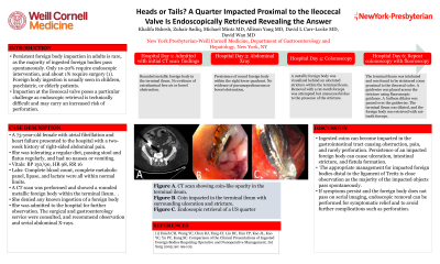
Case Description/Methods: A 73-year-old female with atrial fibrillation and heart failure presented to the hospital with a two week history of right-sided abdominal pain. She was tolerating a regular diet, passing stool and flatus regularly, and had no nausea or vomiting. A CT scan was performed and showed a rounded metallic foreign body within the terminal ileum. The patient denied any known ingestion of a foreign body. She was observed in the hospital for two days with no passage of the foreign body on serial abdominal X-rays. On the third day, endoscopic retrieval was attempted. The terminal ileum was successfully intubated, and a metallic foreign body was visualized behind an ulcerated stricture in the terminal ileum. Removal with a rat-tooth forceps was attempted but was unsuccessful due to the presence of an ileal stricture. Surgery was consulted, but ultimately deferred intervention due to the patient's extensive comorbidities. A repeat colonoscopy was performed. The terminal ileum was intubated and was found to be strictured 10cm proximal to the ileocecal valve. An 0.035 inch guidewire was placed across the stricture using endoscopic and fluoroscopic guidance. A 8-10 mmCRETM Balloon Dilator was passed over the guidewire. The terminal ileum was dilated, and the foreign body was retrieved with rat-tooth forceps. The metallic object was found to be a US quarter. The following day, the patient's pain fully resolved and she was discharged from the hospital.
Discussion: Ingested coins can become impacted in the gastrointestinal tract causing obstruction, pain, and rarely perforation. Persistence of an impacted foreign body can cause ulceration, intestinal stricture, and fistula formation. The appropriate management for impacted foreign bodies distal to the ligament of Treitz is close observation as the majority of the impacted objects pass spontaneously. However, if symptoms persist and the foreign body does not pass on serial imaging, endoscopic removal can be performed for symptomatic relief and to avoid further complications such as perforation.

Disclosures:
Khalifa Bshesh, BA1, Zuhair Sadiq, BA1, Michael J. Mintz, MD2, Allison Yang, MD3, David L. Carr-Locke, MD2, David Wan, MD2. E0286 - Heads or Tails? A Quarter Impacted Proximal to the Ileocecal Valve Is Endoscopically Retrieved Revealing the Answer, ACG 2022 Annual Scientific Meeting Abstracts. Charlotte, NC: American College of Gastroenterology.
1Weill Cornell Medical College, New York, NY; 2New York-Presbyterian Hospital/Weill Cornell Medicine, New York, NY; 3New York Presbyterian Hospital/ Weill Cornell Medicine, New York, NY
Introduction: Persistent foreign body impaction in adults is rare, as the majority of ingested foreign bodies pass spontaneously. Only 10-20% require endoscopic intervention, and about 1% require surgery. Foreign body ingestion is usually seen in children, psychiatric, or elderly patients. Impaction at the ileocecal valve poses a particular challenge as endoscopic retrieval is technically difficult and may carry an increased risk of perforation.
Case Description/Methods: A 73-year-old female with atrial fibrillation and heart failure presented to the hospital with a two week history of right-sided abdominal pain. She was tolerating a regular diet, passing stool and flatus regularly, and had no nausea or vomiting. A CT scan was performed and showed a rounded metallic foreign body within the terminal ileum. The patient denied any known ingestion of a foreign body. She was observed in the hospital for two days with no passage of the foreign body on serial abdominal X-rays. On the third day, endoscopic retrieval was attempted. The terminal ileum was successfully intubated, and a metallic foreign body was visualized behind an ulcerated stricture in the terminal ileum. Removal with a rat-tooth forceps was attempted but was unsuccessful due to the presence of an ileal stricture. Surgery was consulted, but ultimately deferred intervention due to the patient's extensive comorbidities. A repeat colonoscopy was performed. The terminal ileum was intubated and was found to be strictured 10cm proximal to the ileocecal valve. An 0.035 inch guidewire was placed across the stricture using endoscopic and fluoroscopic guidance. A 8-10 mmCRETM Balloon Dilator was passed over the guidewire. The terminal ileum was dilated, and the foreign body was retrieved with rat-tooth forceps. The metallic object was found to be a US quarter. The following day, the patient's pain fully resolved and she was discharged from the hospital.
Discussion: Ingested coins can become impacted in the gastrointestinal tract causing obstruction, pain, and rarely perforation. Persistence of an impacted foreign body can cause ulceration, intestinal stricture, and fistula formation. The appropriate management for impacted foreign bodies distal to the ligament of Treitz is close observation as the majority of the impacted objects pass spontaneously. However, if symptoms persist and the foreign body does not pass on serial imaging, endoscopic removal can be performed for symptomatic relief and to avoid further complications such as perforation.

Figure: Figure 1: A. CT scan showing coin-like opacity in the terminal ileum. B. Coin impacted in the terminal ileum with surrounding ulceration and stricture. C. Endoscopic retrieval of a US quarter
Disclosures:
Khalifa Bshesh indicated no relevant financial relationships.
Zuhair Sadiq indicated no relevant financial relationships.
Michael Mintz indicated no relevant financial relationships.
Allison Yang indicated no relevant financial relationships.
David Carr-Locke indicated no relevant financial relationships.
David Wan indicated no relevant financial relationships.
Khalifa Bshesh, BA1, Zuhair Sadiq, BA1, Michael J. Mintz, MD2, Allison Yang, MD3, David L. Carr-Locke, MD2, David Wan, MD2. E0286 - Heads or Tails? A Quarter Impacted Proximal to the Ileocecal Valve Is Endoscopically Retrieved Revealing the Answer, ACG 2022 Annual Scientific Meeting Abstracts. Charlotte, NC: American College of Gastroenterology.
