Back


Poster Session E - Tuesday Afternoon
Category: GI Bleeding
E0335 - Massive Gastrointestinal Bleed Secondary to Newly Diagnosed Gastric Kaposi Sarcoma
Tuesday, October 25, 2022
3:00 PM – 5:00 PM ET
Location: Crown Ballroom

Has Audio

Spurthi Tarugu, MD
University of Mississippi Medical Center
Jackson, MS
Presenting Author(s)
Spurthi Tarugu, MD, Nathan E. Usry, MD, Zachary Field, MD, Sarah Glover, DO
University of Mississippi Medical Center, Jackson, MS
Introduction: Kaposi sarcoma (KS) is a vascular tumor associated with human herpesvirus 8. It commonly presents with cutaneous lesions but can also have visceral involvement. While presentations of gastrointestinal KS (GI-KS) are highly variable and range from asymptomatic (79%) to nonspecific gastrointestinal symptoms (21%), massive GI hemorrhage is exceedingly rare. Here, we present a case of a massive GI bleed secondary to GI-KS.
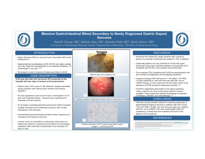
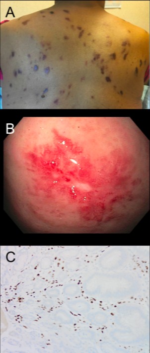
Case Description/Methods: A 39-year-old male with iron deficiency anemia (IDA), HIV/AIDS (CD4 180 cells/mm3), and cutaneous KS compliant with highly-active antiretroviral therapy (HAART) presented to the hospital with two days of melena and hematochezia. On arrival to the emergency room, the patient was hypotensive and with hemoglobin of 4.9 g/dL. Emergent upper endoscopy showed multiple indurated and erythematous lesions in the stomach with central ulceration. Immunohistochemical testing of the biopsy revealed human herpesvirus 8 consistent with Kaposi sarcoma. Lesions were not amenable to endoscopic intervention. The patient was started on liposomal doxorubicin by hematology outpatient.
Discussion: While AIDS-related KS usually presents with cutaneous lesions, it can have visceral involvement in 15% of patients, particularly the gastrointestinal tract, lungs, and lymph nodes. Typically, upper endoscopy is pursued in those who develop GI symptoms (21% of patients) and have a positive occult blood test. The remaining 79% of patients with GI-KS are asymptomatic and are not routinely diagnosed until they develop GI symptoms. We propose that before patients become symptomatic, those who have a CD4 cell count of < 100 cell/μL, HIV RNA >10,000 copies/mLm, men who have sex with men, not on HAART therapy, or have cutaneous KS should be screened with an upper endoscopy as these factors have been shown to be predictive of GI-KS as well as endoscopic severity.
If GI-KS is diagnosed early based on the above screening criteria, patients may have conservative treatment options available. These range from antiretroviral therapy to resection, cryotherapy, radiation, and chemotherapy. Our patient had multiple ulcerated and erythematous lesions on endoscopy, indicative of severe disease not amenable to resection and required chemotherapy. If this patient had been screened when his IDA was discovered, especially given the presence of cutaneous lesions and homosexuality, GI-KS may have been diagnosed and treated with conservative measures prior to its progression and life-threatening presentation.

Disclosures:
Spurthi Tarugu, MD, Nathan E. Usry, MD, Zachary Field, MD, Sarah Glover, DO. E0335 - Massive Gastrointestinal Bleed Secondary to Newly Diagnosed Gastric Kaposi Sarcoma, ACG 2022 Annual Scientific Meeting Abstracts. Charlotte, NC: American College of Gastroenterology.
University of Mississippi Medical Center, Jackson, MS
Introduction: Kaposi sarcoma (KS) is a vascular tumor associated with human herpesvirus 8. It commonly presents with cutaneous lesions but can also have visceral involvement. While presentations of gastrointestinal KS (GI-KS) are highly variable and range from asymptomatic (79%) to nonspecific gastrointestinal symptoms (21%), massive GI hemorrhage is exceedingly rare. Here, we present a case of a massive GI bleed secondary to GI-KS.
Case Description/Methods: A 39-year-old male with iron deficiency anemia (IDA), HIV/AIDS (CD4 180 cells/mm3), and cutaneous KS compliant with highly-active antiretroviral therapy (HAART) presented to the hospital with two days of melena and hematochezia. On arrival to the emergency room, the patient was hypotensive and with hemoglobin of 4.9 g/dL. Emergent upper endoscopy showed multiple indurated and erythematous lesions in the stomach with central ulceration. Immunohistochemical testing of the biopsy revealed human herpesvirus 8 consistent with Kaposi sarcoma. Lesions were not amenable to endoscopic intervention. The patient was started on liposomal doxorubicin by hematology outpatient.
Discussion: While AIDS-related KS usually presents with cutaneous lesions, it can have visceral involvement in 15% of patients, particularly the gastrointestinal tract, lungs, and lymph nodes. Typically, upper endoscopy is pursued in those who develop GI symptoms (21% of patients) and have a positive occult blood test. The remaining 79% of patients with GI-KS are asymptomatic and are not routinely diagnosed until they develop GI symptoms. We propose that before patients become symptomatic, those who have a CD4 cell count of < 100 cell/μL, HIV RNA >10,000 copies/mLm, men who have sex with men, not on HAART therapy, or have cutaneous KS should be screened with an upper endoscopy as these factors have been shown to be predictive of GI-KS as well as endoscopic severity.
If GI-KS is diagnosed early based on the above screening criteria, patients may have conservative treatment options available. These range from antiretroviral therapy to resection, cryotherapy, radiation, and chemotherapy. Our patient had multiple ulcerated and erythematous lesions on endoscopy, indicative of severe disease not amenable to resection and required chemotherapy. If this patient had been screened when his IDA was discovered, especially given the presence of cutaneous lesions and homosexuality, GI-KS may have been diagnosed and treated with conservative measures prior to its progression and life-threatening presentation.

Figure: Figure 1.: (A) KS cutaneous manifestation seen on patient’s back. (B) Multiple indurated, erythematous lesions seen in the gastric body on upper endoscopy. (C) Positive staining for HHV-8 latency-associated nuclear antigen (LNA)-1 confirming the diagnosis of GI-KS.
Disclosures:
Spurthi Tarugu indicated no relevant financial relationships.
Nathan Usry indicated no relevant financial relationships.
Zachary Field indicated no relevant financial relationships.
Sarah Glover: AbbVie – Speakers Bureau. BMS – Consultant. Celgene – Consultant. Janssen – Consultant. Takeda – Speakers Bureau.
Spurthi Tarugu, MD, Nathan E. Usry, MD, Zachary Field, MD, Sarah Glover, DO. E0335 - Massive Gastrointestinal Bleed Secondary to Newly Diagnosed Gastric Kaposi Sarcoma, ACG 2022 Annual Scientific Meeting Abstracts. Charlotte, NC: American College of Gastroenterology.

