Back


Poster Session B - Monday Morning
Category: Liver
B0552 - A Case of Rapidly Progressing Hepatocellular Carcinoma Hidden From Surveillance
Monday, October 24, 2022
10:00 AM – 12:00 PM ET
Location: Crown Ballroom

Has Audio

Jay Shah, DO, MS
Saint Louis University
St. Louis, MO
Presenting Author(s)
Jay Shah, DO, MS, Soumojit Ghosh, MD, Eugene C. Nwankwo, MD, Michelle Baliss, DO, Laith Numan, MD, Hany Elbeshbeshy, MD
Saint Louis University, St. Louis, MO
Introduction: Hepatocellular carcinoma (HCC) accounts for over 90% of all liver cancers worldwide and carries a high mortality rate. Despite screening efforts, the incidence of HCC still remains high due to the prevalence of viral hepatitis, alcoholic cirrhosis, and non alcoholic fatty liver disease. At present, there are no screening guidelines focusing on non cirrhotic primary biliary cholangitis (PBC) patients. We herein present a case of an 84 year old female who was diagnosed with rapidly progressive HCC in the setting of PBC.
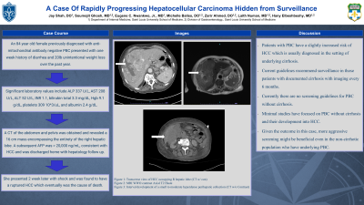
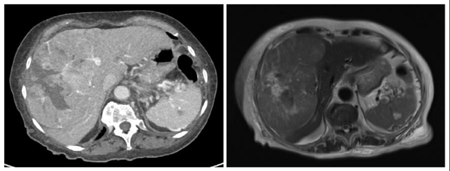
Case Description/Methods: An 84 year old female previously diagnosed with anti-mitochondrial antibody negative PBC presented with one week history of diarrhea and 20lb unintentional weight loss over the past year. Physical exam was notable for non tender hepatomegaly. Significant laboratory values include ALP 337 U/L, AST 208 U/L, ALT 52 U/L, INR 1.1, bilirubin total 3.3 mg/dL, Hgb 9.1 g/dL, platelets 309 10^3/uL, and albumin 2.4 g/dL. Social history was negative for tobacco or alcohol use. A CT of the abdomen and pelvis was obtained and revealed a 16 cm mass encompassing the entirety of the right hepatic lobe. A subsequent AFP was > 20,000 ng/mL, consistent with HCC and was discharged home with hepatology follow up. She presented 2 week later with shock and was found to have a ruptured HCC which eventually was the cause of death. 6 months prior to admission, she was noted to have normal liver enzymes and AFP < 2 ng/mL, but no imaging was obtained as a fibroscan done 12 months prior to admission revealed a liver stiffness of 7.8 kPa (low to moderate probability of advanced fibrosis) and CAP of 229 dB/m (no liver steatosis).
Discussion: Patients with PBC have a slightly increased risk of HCC which is usually diagnosed in the setting of underlying cirrhosis. Current guidelines recommend surveillance in those patients with documented cirrhosis with imaging every 6 months. However, studies have shown that patients with PBC have a 2.4% risk of developing HCC at any histologic stage of PBC, with a male predominance. There are currently no screening guidelines for PBC, which may have led to an earlier diagnosis, and therefore treatment, in our patient who passed away only 2 weeks after presentation due to ruptured HCC. Given the outcome in this case, more aggressive screening might be beneficial even in the non-cirrhotic population who have underlying PBC.

Disclosures:
Jay Shah, DO, MS, Soumojit Ghosh, MD, Eugene C. Nwankwo, MD, Michelle Baliss, DO, Laith Numan, MD, Hany Elbeshbeshy, MD. B0552 - A Case of Rapidly Progressing Hepatocellular Carcinoma Hidden From Surveillance, ACG 2022 Annual Scientific Meeting Abstracts. Charlotte, NC: American College of Gastroenterology.
Saint Louis University, St. Louis, MO
Introduction: Hepatocellular carcinoma (HCC) accounts for over 90% of all liver cancers worldwide and carries a high mortality rate. Despite screening efforts, the incidence of HCC still remains high due to the prevalence of viral hepatitis, alcoholic cirrhosis, and non alcoholic fatty liver disease. At present, there are no screening guidelines focusing on non cirrhotic primary biliary cholangitis (PBC) patients. We herein present a case of an 84 year old female who was diagnosed with rapidly progressive HCC in the setting of PBC.
Case Description/Methods: An 84 year old female previously diagnosed with anti-mitochondrial antibody negative PBC presented with one week history of diarrhea and 20lb unintentional weight loss over the past year. Physical exam was notable for non tender hepatomegaly. Significant laboratory values include ALP 337 U/L, AST 208 U/L, ALT 52 U/L, INR 1.1, bilirubin total 3.3 mg/dL, Hgb 9.1 g/dL, platelets 309 10^3/uL, and albumin 2.4 g/dL. Social history was negative for tobacco or alcohol use. A CT of the abdomen and pelvis was obtained and revealed a 16 cm mass encompassing the entirety of the right hepatic lobe. A subsequent AFP was > 20,000 ng/mL, consistent with HCC and was discharged home with hepatology follow up. She presented 2 week later with shock and was found to have a ruptured HCC which eventually was the cause of death. 6 months prior to admission, she was noted to have normal liver enzymes and AFP < 2 ng/mL, but no imaging was obtained as a fibroscan done 12 months prior to admission revealed a liver stiffness of 7.8 kPa (low to moderate probability of advanced fibrosis) and CAP of 229 dB/m (no liver steatosis).
Discussion: Patients with PBC have a slightly increased risk of HCC which is usually diagnosed in the setting of underlying cirrhosis. Current guidelines recommend surveillance in those patients with documented cirrhosis with imaging every 6 months. However, studies have shown that patients with PBC have a 2.4% risk of developing HCC at any histologic stage of PBC, with a male predominance. There are currently no screening guidelines for PBC, which may have led to an earlier diagnosis, and therefore treatment, in our patient who passed away only 2 weeks after presentation due to ruptured HCC. Given the outcome in this case, more aggressive screening might be beneficial even in the non-cirrhotic population who have underlying PBC.

Figure: CT and MRI visualizing HCC predominately in right hepatic lobe
Disclosures:
Jay Shah indicated no relevant financial relationships.
Soumojit Ghosh indicated no relevant financial relationships.
Eugene Nwankwo indicated no relevant financial relationships.
Michelle Baliss indicated no relevant financial relationships.
Laith Numan indicated no relevant financial relationships.
Hany Elbeshbeshy indicated no relevant financial relationships.
Jay Shah, DO, MS, Soumojit Ghosh, MD, Eugene C. Nwankwo, MD, Michelle Baliss, DO, Laith Numan, MD, Hany Elbeshbeshy, MD. B0552 - A Case of Rapidly Progressing Hepatocellular Carcinoma Hidden From Surveillance, ACG 2022 Annual Scientific Meeting Abstracts. Charlotte, NC: American College of Gastroenterology.

